



胆道がん(胆管がん・胆嚢がん)について

- 胆道ガン(胆嚢がん、胆管がん)とは
- 胆道ガン(胆嚢がん、胆管がん)の症状
- 胆道ガン(胆嚢がん、胆管がん)の原因
- 胆道ガン(胆嚢がん、胆管がん)の検査方法や診断
- 胆道ガン(胆嚢がん、胆管がん)の治療方法
- 胆道ガン(胆嚢がん、胆管がん)の予防方法
- 胆道ガン(胆嚢がん、胆管がん)の患者さんによる体験談
- まとめ
胆道ガンは、70歳代・80歳代の発症率がとても多いと言われているがんです。
年齢とともに発症率や死亡率が上がるのが特徴で、胆管がんは男性が、胆嚢がんは女性の患者さんが多いです。
初期症状が現れにくく、広範囲にがん細胞が広がりやすい特徴から、病状が進行してから発見されるケースが多いがんです。
今回は、そんな胆道ガンについて詳しく解説していきたいと思います。
胆道ガン(胆嚢がん、胆管がん)とは
それでは早速、胆道ガン(胆嚢がん、胆管がん)とはどのような疾患なのかを解説していきたいと思います。
私たちの肝臓から分泌された胆汁は、胆管を通り一時的に胆嚢にためられます。
そしてその後、乳頭という出口から十二指腸へと流れるのですが、この十二指腸へ流れる胆汁の通り道を「胆道」と言います。
この胆道の各部位に発生する悪性腫瘍の事を、まとめて胆道ガンと呼びます。
胆道ガンは、胆嚢がんと胆管がんをまとめた総称でもあり、2つに分けて解説される事が多いと言われています。
また胆道ガンは、胆道が十二指腸に繋がっている事や、胆管の壁が薄い事、近くに重要な血管や細かい神経が通っていたり、リンパ節が豊富に存在している事などから、臓器の構造上がん細胞が広範囲に及びやすいという特徴もあります。
このような理由から、胆道ガンは、診断が確定した時にはかなり病状が進行しているという事も少なくないのです。
胆道ガン(胆嚢がん、胆管がん)の症状
では次に、胆道ガン(胆嚢がん、胆管がん)の症状について解説していきましょう。
胆道ガンは、初期の段階では症状がほとんど現れない事が多いがんです。
そのため、ある程度がんが進行するまでは、無症状で過ごしてしまう事が多いと言われています。
偶然、健康診断の腹部超音波検査や、胆石症を伴う胆嚢摘出術の際に、偶然発見される事もありますが稀でしょう。
胆道ガンが進行してくると、「黄疸」「みぞおちや右の脇腹の痛み」「吐き気」「嘔吐」「体重減少」のような症状が現れてきます。
この時現れる黄疸は、腫瘍が大きくなって胆嚢管や胆管が狭くなり、胆汁が血液中に流れ出てくるため現れるものです。
黄疸がさらに悪化してくると、「便が白っぽくなってくる」「尿の色が茶色く濃くなる」「皮膚のかゆみが出てくる」というような症状が現れてきます。
このような黄疸を機に見つかるものは、すでに胆道ガンがかなり進行している状態と言えるでしょう。
胆道ガン(胆嚢がん、胆管がん)の原因
では次に、どのような原因で胆道ガンが発生するのかを解説していきたいと思います。
胆道ガンの発症原因は、現在のところ解明されていないのが現状です。
しかし、胆道に長期的に炎症や刺激が続く事で、胆道ガンの発症に繋がるのではないかと言われています。
その中でも、特に具体的な危険因子として挙げられるのが、胆嚢がん・胆管がんともに、膵・胆管合流異常があります。
膵・胆管合流異常は、膵管と胆管が十二指腸に入る手前で、合流してしまう先天性の形成異常です。
膵液や胆汁が相互に逆流してしまう事で、胆道ガンをはじめ様々な病態を引き起こすと言われているのです。
また胆管がんは、発症の9割以上が結石のある部位に集中している事から、肝内結石症との関連性も多く報告されています。
胆道ガン(胆嚢がん、胆管がん)の検査方法や診断
では次に、胆道ガン(胆嚢がん、胆管がん)の検査方法や診断について解説していきましょう。
胆道ガンは、まず血液検査によって、肝・胆道系酵素や血清ビリルビンの濃度が高値になっていないかを確認します。
しかし、肝・胆道系酵素や血清ビリルビンの濃度は、必ずしも全て胆道ガンで上昇するとは限らない為、補助的な検査となっています。
そのため、さらに血液検査と腹部超音波検査を併用して行います。
腹部超音波検査では、胆管が拡張していないか、胆のうの壁が厚くなっていないか、盛り上がっていないかなどを調べ病変を確認していきます。
そして、この時に病変を発見した場合は、部位や目的に応じて、CT検査やMRI検査などによって、より高度な検査を実施していきます。
さらに、十二指腸に内視鏡を挿入して、胆管と膵管に造影剤を入れて、X線撮影をする「内視鏡的逆行性胆管膵管造影(ERCP)」、先端に超音波装置が付いた内視鏡を胆嚢付近の胃や十二指腸に挿入して体内で超音波検査を行う「超音波内視鏡検査(EUS)」などを行う事もあります。
さらにPET検査では、主にリンパ節への転移や遠隔転移の有無を調べます。
胆道ガン(胆嚢がん、胆管がん)の治療方法
では次に、胆道ガン(胆嚢がん、胆管がん)の治療方法について解説していきたいと思います。
胆道ガンの治療は、外科的手術によってがん細胞を取り除く事が中心となります。
手術の方法は、がんが発生している部位によって異なりますが、胆管とともに膵臓と十二指腸を切除したり、肝臓も切除するケースもあります。
また、場合によっては膵臓と肝臓の両方を切除しなければならない事もあります。
門脈・肝動脈などの重要血管にがんが広がっている場合は、切除後に再建の必要があります。
肝臓や肺・骨への転移・腹膜へのがんの広がり・遠隔リンパ節転移を伴う胆道ガンの場合は、切除術は推奨されておらず手術が難しくなります。
そのようなケースでは、抗がん剤による化学療法や放射線治療を行います。
また、黄疸による肝不全を防ぐために、胆嚢にたまった胆汁を取り除く胆道ドレナージや、内視鏡を用いて胆管内にチューブや金属製の器具を留置して胆汁の流れを確保する胆道ステントを行う事もあります。
胆道ガン(胆嚢がん、胆管がん)の予防方法
では次に、胆道ガン(胆嚢がん、胆管がん)の予防方法について解説していきましょう。
胆道ガンの大きな発症原因としては、膵・胆管合流異常が挙げられるので、膵・胆管合流異常と診断されたら、予防的に手術で胆嚢や拡張した胆管を摘出する事が予防に繋がります。
また、膵・胆管合流異常と同じく、胆道ガンの発症危険因子として挙げられる胆石症も、予防的に胆嚢摘出手術を行う事で予防に繋がるでしょう。
その他には、定期的に超音波検査を受けて、胆嚢に異常がないか把握しておく事も大切です。
胆道ガン(胆嚢がん、胆管がん)の患者さんによる体験談
それでは最後に、胆道ガン(胆嚢がん、胆管がん)の患者さんによる体験談をご紹介していきたいと思います。
70歳代 女性
「久しぶりに体重を測ってみると、体重が減少していて食欲不振が続くようになっていました。
最初は、体調が悪いのだろうと思っていたのですが、なかなか回復しないので心配になり病院を受診しました。
そこで、腹部のエコー検査を行ったところ、肝臓に8センチもの腫瘍があるのが見つかったのです。
すぐに総合病院で詳しい検査をしたところ、胆管がんで肺転移、リンパ節転移、肝臓にも複数の転移がある状態でした。
あまりの転移の多さに、摘出手術は難しいという事になり、抗がん剤による化学療法で治療をしています。
その効果か、がん細胞が小さくなってきていています。まだ転移の危険性はあるものの少しでも長生きしたいと思っています。」
まとめ
今回は、胆道ガン(胆嚢がん、胆管がん)について詳しく解説してきました。
胆道ガン(胆嚢がん、胆管がん)は、がんの中でも特に初期症状が現れにくい疾患で、診断が確定した時点では病状がかなり進行している事も珍しくありません。
そのため、定期的な検査や、少しでも体調に異変を感じたら、すぐに病院を受診する事が早期に発見する為に必要なのです。
先天性の異常による原因が、ほとんどと言われている胆道ガン(胆嚢がん、胆管がん)。
発症に繋がる病気の治療をしっかりと行うことで予防に繋がるため、少しでもサインがあれば放置せずに病院に行きましょう。
胆道癌 胆管癌 胆嚢癌
==============================
Biliary tract cancer is a cancer that is said to have a very high incidence in the 70s and 80s. The
It is characterized by an increase in incidence and mortality with age, with many male patients having bile duct cancer and female patients having gallbladder cancer. The
It is a cancer that is often found after the condition has progressed because the initial symptoms are less likely to appear and the cancer cells tend to spread over a wide area. The
This time, I would like to explain in detail about such biliary tract cancer. T
Heading 1, what is biliary tract cancer?
The
Now, I would like to explain what kind of disease is biliary tract cancer (gallbladder cancer, bile duct cancer). The
The
Bile secreted by our liver passes through the bile ducts and is temporarily stored in the gallbladder. The
After that, it flows from the exit called the nipple to the duodenum, and the path of bile that flows to the duodenum is called the "bile duct". The
The malignant tumors that occur in each part of the biliary tract are collectively called biliary tract cancer. The
The
Biliary tract cancer is also a general term for gallbladder cancer and bile duct cancer, and is often explained in two parts. The
In addition, biliary tract cancer is an organ because the biliary tract is connected to the duodenum, the wall of the bile duct is thin, important blood vessels and small nerves pass nearby, and lymph nodes are abundant. It is also structurally easy for cancer cells to spread over a wide area. The
The
For this reason, biliary tract cancer is often quite advanced when the diagnosis is confirmed. The
The
Heading 2, symptoms of biliary tract cancer
The
Next, let's explain the symptoms of biliary tract cancer. The
The
Biliary tract cancer is a cancer that often has few symptoms in the early stages. The
Therefore, it is said that people often spend their time asymptomatic until the cancer progresses to some extent. The
It may be discovered by chance during an abdominal ultrasonography for a medical examination or a cholecystectomy with cholecystectomy, but it is rare. The
The
As biliary tract cancer progresses, symptoms such as "jaundice," "epigastrium and right flank pain," "nausea," "vomiting," and "weight loss" appear. The
Jaundice that appears at this time appears because the tumor grows larger, the cystic ducts and bile ducts narrow, and bile flows out into the blood. The
The
As jaundice worsens, symptoms such as "feces become whitish," "urine becomes brown and dark," and "skin itching appears." The
If you find such jaundice as an opportunity, it can be said that biliary tract cancer has already progressed considerably. The
The
Heading 3, Causes of Biliary Cancer
The
Next, I would like to explain what causes biliary tract cancer. The
The
At present, the cause of biliary tract cancer has not been clarified. The
However, it is said that long-term inflammation and irritation of the biliary tract may lead to the development of biliary tract cancer. The
The
Among them, the most specific risk factors are pancreatic and bile duct confluence abnormalities in both gallbladder cancer and bile duct cancer. The
Pancreatic-bile duct confluence is a congenital dysplasia that merges before the pancreatic duct and bile duct enter the duodenum. The
It is said that pancreatic juice and bile flow back to each other, causing various pathological conditions including biliary tract cancer. The
The
In addition, since more than 90% of the onset of bile duct cancer is concentrated in the area with stones, many reports have been made regarding the association with intrahepatic stone disease. The
The
Heading 4, examination method and diagnosis of biliary tract cancer
The
Next, let's explain how to test and diagnose biliary tract cancer. The
The
For biliary tract cancer, a blood test is first performed to check for high levels of liver / biliary enzymes and serum bilirubin. The
However, the concentrations of liver / biliary enzymes and serum bilirubin do not always increase in biliary tract cancer, so this is a supplementary test. The
Therefore, a blood test and an abdominal ultrasonography will be performed together. The
The
In abdominal ultrasonography, the bile duct is not dilated, the wall of the gallbladder is not thickened, or it is raised, and the lesion is confirmed. The
If a lesion is found at this time, more advanced examinations will be performed by CT examination or MRI examination, depending on the site and purpose. The
In addition, "endoscopic retrograde cholangiopancreatography (ERCP)", in which an endoscope is inserted into the duodenum, a contrast agent is put into the bile duct and pancreatic duct, and X-rays are taken, and an ultrasonic device is attached to the tip. "Endoscopic ultrasonography (EUS)", in which an endoscope is inserted into the stomach or duodenum near the gallbladder and an ultrasonic examination is performed inside the body, may be performed. The
In addition, PET scans mainly look for metastases to lymph nodes or distant metastases. The
The
Heading 5, treatment method for biliary tract cancer
The
Next, I would like to explain how to treat biliary tract cancer. The
The
Treatment of biliary tract cancer is centered on removing cancer cells by surgery. The
The method of surgery depends on where the cancer is occurring, but in some cases, the pancreas and duodenum are removed along with the bile duct, and the liver is also removed. The
In some cases, both the pancreas and liver may need to be removed. The
The
If the cancer has spread to important blood vessels such as the portal vein and hepatic artery, it needs to be reconstructed after excision. The
The
For biliary tract cancer with metastasis to the liver, lungs, bones, spread of cancer to the peritoneum, or distant lymph node metastasis, resection is not recommended and surgery is difficult. The
In such cases, chemotherapy or radiation therapy with anticancer drugs is given. The
The
In addition, in order to prevent liver failure due to jaundice, biliary drainage to remove bile accumulated in the gallbladder and biliary stenting to secure the flow of bile by placing a tube or metal instrument in the bile duct using an endoscope are performed. There are also things. The
The
Heading 6, how to prevent biliary tract cancer
The
Next, let's explain how to prevent biliary tract cancer. The
The
Since pancreatic / bile duct abnormalities are a major cause of biliary tract cancer, if pancreatic / bile duct abnormalities are diagnosed, prophylactic surgery to remove the gallbladder and dilated bile ducts will lead to prevention. The
In addition, cholecystectomy, which is one of the risk factors for developing biliary tract cancer, can be prevented by performing prophylactic cholecystectomy, as is the case with abnormal pancreatic and bile duct confluence. The
In addition, it is important to have regular ultrasound examinations to find out if there are any abnormalities in the gallbladder.
Heading 7, experiences by patients with biliary tract cancer
The
Finally, I would like to introduce the experiences of patients with biliary tract cancer. The
The
Female in her 70s
"When I weighed myself for the first time in a while, I found that I was losing weight and had anorexia.
At first, I thought she was sick, but she was worried because she couldn't recover easily, so she went to the hospital. The
She performed an echography of her abdomen and found a tumor as large as 8 centimeters in her liver. The
She soon had a detailed examination at a general hospital where she had bile duct cancer with lung metastases, lymph node metastases, and multiple metastases to the liver. The
Due to the large number of metastases, excision surgery is difficult, and she is treated with chemotherapy with anticancer drugs. The
Perhaps because of that effect, the cancer cells are getting smaller. Although there is still a risk of metastasis, I want to live a little longer. 』
The
Heading 8, Summary
The
This time, I have explained in detail about biliary tract cancer. The
Biliary tract cancer is a disease in which the initial symptoms are particularly unlikely to appear, and it is not uncommon for the condition to progress considerably when the diagnosis is confirmed. The
Therefore, it is necessary to have regular examinations and to see a hospital immediately if you feel any abnormalities in your physical condition in order to detect it at an early stage. The
Biliary tract cancer (gallbladder cancer, bile duct cancer) is said to be the most common cause of congenital anomalies. The
Proper treatment of the illness that leads to the onset will lead to prevention, so if there is any sign, go to the hospital without leaving it.
すい臓がんについて About Pancreatic cancer
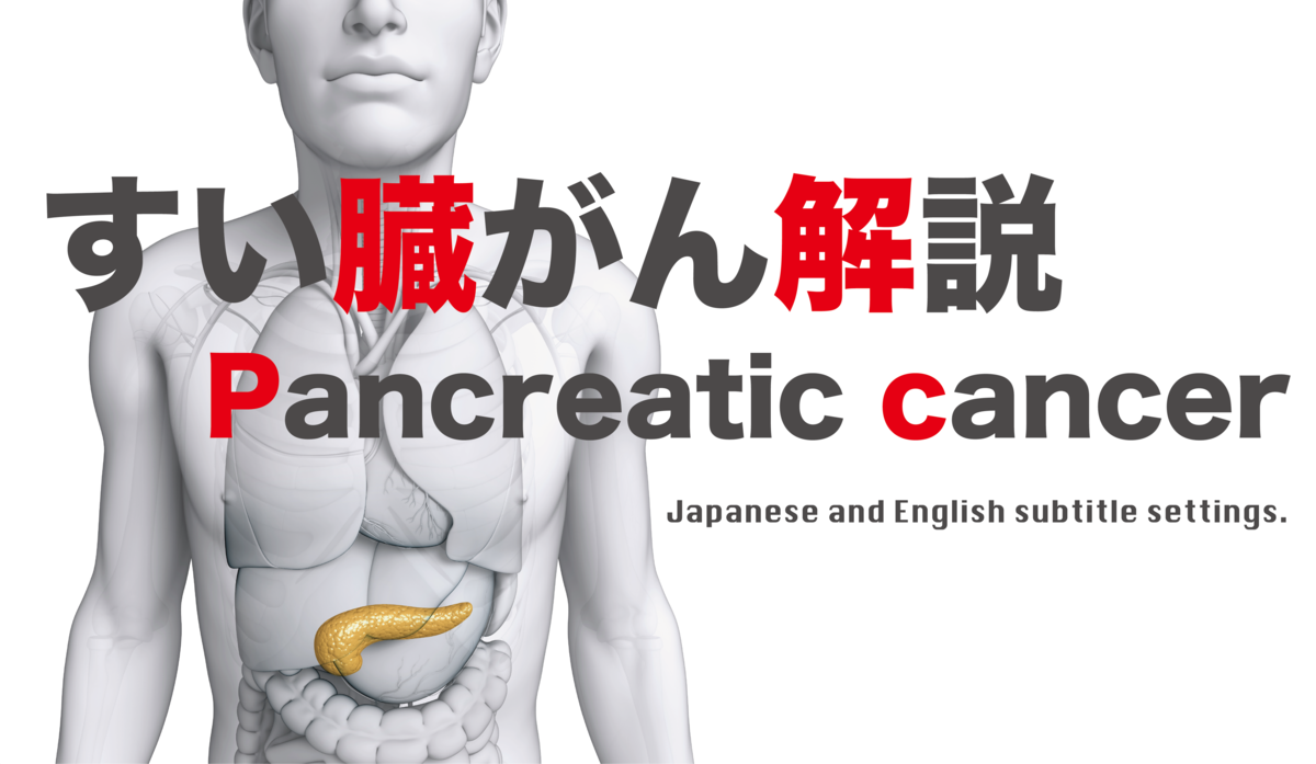
- すい臓がんとは
- すい臓がんの原因
- すい臓がんの症状
- すい臓がんの検査
- すい臓がんの治療
- すい臓がんの予防方法
- すい臓がんの生存率
- すい臓がん患者の体験談
- まとめ
- What is pancreatic cancer?
- Causes of pancreatic cancer
- Symptoms of pancreatic cancer
- Pancreatic cancer test
- Treatment of pancreatic cancer
- How to prevent pancreatic cancer
- Survival rate of pancreatic cancer
- Experiences of pancreatic cancer patients
- summary
すい臓がんは、21世紀に残された最後の消化器がんと言われているほど、課題の多いがんでもあります。
国立がん研究センターのがん統計によると、すい臓がんの患者数は男女合わせて1年間で約3万3,000人となっています。
患者数は、がんの中では少ない方ですが、年間死亡数は約3万にのぼり、肺がんや胃がんに次いで第5位となり、生存率もとても低い特徴があります。
今回は、そんなすい臓がんについて詳しく解説していきたいと思います。
すい臓がんとは
それでは早速、すい臓がんがどのようながんなのかを解説いていきましょう。
すい臓がんは、すい臓から発生する悪性腫瘍で、9割以上がすい臓から十二指腸に分泌される消化液の通り道である膵管に出来ます。
このため、一般的にすい臓がんとは膵管がんの事を指します。
すい臓がんは、すい臓が他の臓器に囲まれているので、腫瘍が見つけにくく組織採取も難しいという特徴があります。
また、転移のスピードも速く早期の段階でリンパ管などに広がります。
約7割が手術での治療が不可能という難しいがんで、再発の可能性が高い傾向があります。
平均5年生存率は20~40%ととても低く、年齢が上がるにつれて発症率が高くなるがんです。
すい臓がんの原因
それでは次に、すい臓がんの原因は、どのようなものが考えられるのでしょうか。
すい臓がんの原因は、主にライフスタイルや遺伝性が強いと言われています。
また、慢性膵炎などがすい臓がんのリスク要因になる事も多いとされています。
慢性膵炎は、習慣的に多量のアルコールを摂取していたり、慢性的に強いストレスにさらされている人に多く発症します。
遺伝性に関しては、親や兄弟姉妹など血縁関係者の中に、すい臓がん患者が2人以上いる場合、家族性すい臓がんの診断がされます。
また、脂肪分の多い食事を摂る人や、糖尿病にかかっている人はすい臓がんの発症率が高いという研究データも出ています。
糖尿病と診断された時は、一緒にすい臓がんの検査を受けることが推奨されているほど関連が強く、急激に血糖コントロールが悪化したした場合などすい臓がんが隠れているという事も多いのです。
すい臓がんの症状
すい臓がんは、初期症状がほとんどないのが特徴です。
自覚症状が出てくる頃には、がんがかなり進行している事がほとんどと言われています。
すい臓がんは、進行するにつれて「腹部膨満感」「食欲不振」「体重減少」「全身の倦怠感」などが症状として現れてきます。
また、膵頭部にがんが発生した場合は、がんが胆管を圧迫する為、胆汁の流れが悪くなり黄疸の症状が現れます。
すい臓がんの検査
すい臓がんの診断をする為の検査としては、「血液検査で腫瘍マーカーの確認」「腹部超音波検査」「CT検査」「MRI検査」「超音波内視鏡検査」などがあります。
超音波内視鏡検査は、2cm以下の小さな腫瘍も比較的見つけやすく、先端に小さな超音波のついた内視鏡を挿入する事でがんを見つける検査です。
第1段階の検査で超音波内視鏡検査を行い、そこで診断が難しいと判断された場合は、他の検査と併せて詳しく調べていきます。
例えば、内視鏡の先端から胆管・膵管にカテーテルを入れて、造影検査を行う内視鏡的逆行性胆道膵管造影や、放射性の薬剤を使って全身へのがん細胞の広がりを調べるPET検査などがあります。
また生検と言って、内視鏡や腹腔鏡で腫瘍の一部を採取し、顕微鏡で観察する検査を行う事もあります。
このように様々な検査を組み合わせ、その検査結果を総合的に判断して診断されます。
すい臓がんの治療
それでは次に、すい臓がんの治療について解説していきたいと思います。
すい臓がんの治療には、主に「手術」「抗がん剤による化学療法」「放射線治療」の3つがあります。
これらの治療法は、検査結果によってがんの進行度合いや、患者の年齢・体の状態に合わせて選択されます。
がんが比較的初期の段階で、がん細胞がすい臓周辺の臓器へ転移していない場合は、手術と化学療法を組み合わせて治療するのが一般的です。
しかし、すい臓がんは初期症状があまり出ない為、すい臓がんの診断がされた時点で、ほとんどの場合はがんが進行している事が多く、すい臓周辺の臓器へ転移している事が多いです。
すい臓がんを手術で治療する場合は、すい臓の一部と十二指腸、胆管、胆のうを切除する方法と、すい臓の一部と脾臓をまとめて取り除く方法、すい臓をすべて切除する方法があります。
どの手術内容にするかは、がんが発生している位置や広がりを確認して選択します。
すい臓がんが進行していて、他の臓器への転移がある場合は手術が出来ない為、化学療法や放射線治療を行う事になります。
すい臓がんの予防方法
では次に、すい臓がんを予防する方法をご紹介したいと思います。
すい臓がんを予防する為には、発生因子と言われている膵炎を防ぐ予防方法が効果的です。
過度の飲酒、肉類を中心とした脂質の多い食事、カフェインの多量接種、香辛料や刺激物の過剰摂取は、膵炎の発生を誘発させるので避けたいものです。
また、糖尿病との関連もすい臓がんにはあるため、肥満を防ぐ為の野菜中心のバランスの良い食事や生活習慣病の予防も効果的です。
さらに、既に糖尿病や慢性膵炎を患っている人は、定期的に医療機関で検査を受けることが予防方法として有効です。
特に、腹部に違和感がある場合は早めに病院に行きましょう。
そして、すい臓がんだけに限らず、がん全体に言えることですが喫煙はがんの要因となり得ますので、禁煙を推奨しています。
すい臓がんの生存率
すい臓がんは、がんの中でも代表的な難治がんで、生存率も極めて低い特徴があります。
比較的初期の段階でがんを発見でき、手術出来た人の術後5年生存率は10~30%となっています。
そして、他の臓器への転移はないが手術ができない人の場合の1年生存率は30~50%。
他の臓器への転移があり手術が出来ない人の場合は、1年生存率は10~30%となっています。
すい臓がん患者の体験談
それでは最後に、すい臓がん患者の体験談をご紹介していきましょう。
49歳 男性
「平成17年に、進行性のすい臓がんの告知を受けました。週1回の抗がん剤投与と、所要時間30分程度の放射線療法を毎日6週間続けました。
退院後は、2週に1回のペースで抗がん剤治療を続け、結果約1年半後には腫瘍マーカーも下がりました。
2年目の春に体重が減り始め、一進一退の状態が続いています。」
51歳 男性
「50歳の誕生日を迎え、会社の検診結果が全て良好だった翌月、突然オレンジ色の尿と全身の痒みが襲ってきました。
病院での診察の結果、すい臓に2cmほどの塊があり、膵管と胆管が押しつぶされている事が分かりました。
すぐに紹介状をもらって大きな病院に行き、入院しすい臓を切除しました。ちょうど術後1年の時に、肺への転移が見つかり、本格的な抗がん剤治療が始まりました。
まだまだ抗がん剤治療は続きますが、悔いの残らないように過ごしていきたいと思っています。」
まとめ
さて今回は、がんの中でも難治として知られている「すい臓がん」について解説してきました。
すい臓がんは、初期症状が出にくく診断された時には、すでに進行している場合がほとんどです。
そのため、普段から生活習慣に気をつけ、定期的な検診を受けることで可能な限り防ぐ事が大切なのです。
=======================================================
Pancreatic cancer is also a cancer with many challenges, as it is said to be the last gastrointestinal cancer left in the 21st century.
According to the Cancer Statistics of the National Cancer Center, the total number of patients with pancreatic cancer is about 33,000 per year for both men and women.
Although the number of patients is small among cancers, the annual number of deaths is about 30,000, which is the fifth highest after lung cancer and stomach cancer, and the survival rate is also very low.
This time, I would like to explain in detail about such pancreatic cancer.
What is pancreatic cancer?
Now let's explain what kind of cancer pancreatic cancer is.
Pancreatic cancer is a malignant tumor that develops in the pancreas, and more than 90% of it forms in the pancreatic duct, which is the path of digestive juice secreted from the pancreas into the duodenum.
For this reason, pancreatic cancer generally refers to pancreatic duct cancer.
Pancreatic cancer is characterized by the fact that the pancreas is surrounded by other organs, making it difficult to find the tumor and collect tissue.
In addition, the speed of metastasis is fast and spreads to lymphatic vessels at an early stage.
About 70% have difficult cancers that cannot be treated by surgery, and there is a high possibility of recurrence.
The average 5-year survival rate is as low as 20-40%, and the incidence increases with age.
Causes of pancreatic cancer
Then, what are the possible causes of pancreatic cancer?
It is said that the causes of pancreatic cancer are mainly lifestyle and heredity.
In addition, it is said that chronic pancreatitis is often a risk factor for pancreatic cancer.
Chronic pancreatitis often occurs in people who are habitually ingesting large amounts of alcohol or who are chronically exposed to intense stress.
With regard to heritage, familial pancreatic cancer is diagnosed when there are two or more patients with pancreatic cancer among relatives such as parents and siblings.
Studies have also shown that people who eat a fatty diet or who have diabetes have a higher incidence of pancreatic cancer.
When a person is diagnosed with diabetes, the relationship is so strong that it is recommended to have a pancreatic cancer test together, and pancreatic cancer is often hidden when blood sugar control suddenly deteriorates. is.
Symptoms of pancreatic cancer
Pancreatic cancer is characterized by few initial symptoms.
By the time subjective symptoms appear, it is said that most of the cancer has progressed considerably.
As pancreatic cancer progresses, symptoms such as "abdominal bloating," "loss of appetite," "weight loss," and "general malaise" appear.
In addition, when cancer develops in the head of the pancreas, the cancer presses on the bile ducts, resulting in poor bile flow and jaundice symptoms.
Pancreatic cancer test
Examinations for diagnosing pancreatic cancer include "confirmation of tumor markers by blood test", "abdominal ultrasonography", "CT examination", "MRI examination", and "endoscopic ultrasonography".
Endoscopic ultrasonography is a test that finds cancer by inserting an endoscope with a small ultrasound at the tip, which makes it relatively easy to find small tumors of 2 cm or less.
Endoscopic ultrasonography is performed in the first stage of the examination, and if it is determined that the diagnosis is difficult, we will investigate it in detail together with other examinations.
For example, endoscopic retrograde cholangiopancreatography, in which a catheter is inserted into the bile duct / pancreatic duct from the tip of the endoscope to perform contrast examination, and PET examination, which examines the spread of cancer cells throughout the body using a radiopharmaceutical. And so on.
In addition, a biopsy may be performed by taking a part of the tumor with an endoscope or laparoscope and observing it with a microscope.
In this way, various tests are combined and the test results are comprehensively judged for diagnosis.
Treatment of pancreatic cancer
Next, I would like to explain the treatment of pancreatic cancer.
There are three main types of treatment for pancreatic cancer: surgery, chemotherapy with anticancer drugs, and radiation therapy.
These treatments are selected according to the degree of cancer progression and the patient's age and physical condition, depending on the test results.
If the cancer is relatively early and the cancer cells have not spread to the organs around the pancreas, it is common to treat it with a combination of surgery and chemotherapy.
However, since pancreatic cancer does not have many initial symptoms, in most cases the cancer has progressed at the time of diagnosis of pancreatic cancer, and it has spread to the organs around the pancreas. There are many.
When treating pancreatic cancer by surgery, there are two methods: removing part of the pancreas and duodenum, bile duct, and gallbladder, removing part of the pancreas and spleen together, and removing the entire pancreas.
The surgery content should be selected by checking the location and spread of the cancer.
If pancreatic cancer is progressing and has metastasized to other organs, surgery cannot be performed, so chemotherapy or radiation therapy will be required.
How to prevent pancreatic cancer
Next, I would like to introduce how to prevent pancreatic cancer.
In order to prevent pancreatic cancer, a preventive method to prevent pancreatitis, which is said to be a causative factor, is effective.
Excessive drinking, a high-fat diet centered on meat, high doses of caffeine, and excessive intake of spices and stimulants can induce pancreatitis and should be avoided.
In addition, since pancreatic cancer is also associated with diabetes, it is effective to have a well-balanced diet centered on vegetables and prevent lifestyle-related diseases to prevent obesity.
In addition, for people who already have diabetes or chronic pancreatitis, regular medical examinations are an effective preventive measure.
In particular, if you feel something is wrong with your abdomen, go to the hospital as soon as possible.
And, not only for pancreatic cancer, but for cancer as a whole, smoking can be a factor in cancer, so we recommend that you quit smoking.
Survival rate of pancreatic cancer
Pancreatic cancer is a typical intractable cancer and has an extremely low survival rate.
The 5-year survival rate after surgery is 10 to 30% for those who can detect cancer at a relatively early stage and can undergo surgery.
The one-year survival rate for people who have not metastasized to other organs but cannot be operated on is 30 to 50%.
For people who have metastases to other organs and cannot be operated on, the 1-year survival rate is 10 to 30%.
Experiences of pancreatic cancer patients
Finally, let me introduce the experiences of pancreatic cancer patients.
49-year-old man
"In 2005, I was notified of advanced pancreatic cancer. I continued to receive anti-cancer drugs once a week and radiation therapy for about 30 minutes every day for 6 weeks.
After discharge, he continued to receive chemotherapy once every two weeks, resulting in a decrease in tumor markers about a year and a half later.
In the spring of the second year, I started to lose weight, and I'm still in a state of progress and retreat. "
51-year-old man
"On his 50th birthday, the month after the company's screenings were all good, suddenly orange urine and generalized itching struck.
A medical examination at the hospital revealed that the pancreas had a mass of about 2 cm and the pancreatic duct and bile duct were crushed.
He immediately received a letter of introduction and went to a large hospital where he was hospitalized and had his pancreas removed. Just one year after the operation, metastasis to the lung was found and full-scale anticancer drug treatment began.
Anti-cancer drug treatment will continue, but I would like to spend my time without regrets. "
summary
This time, I have explained about "pancreatic cancer," which is known to be intractable among cancers.
In most cases, pancreatic cancer has already progressed when the initial symptoms are difficult to develop and it is diagnosed.
Therefore, it is important to pay attention to your lifestyle and to prevent it as much as possible by undergoing regular medical examinations.
子宮がんについて About Uterine cancer.
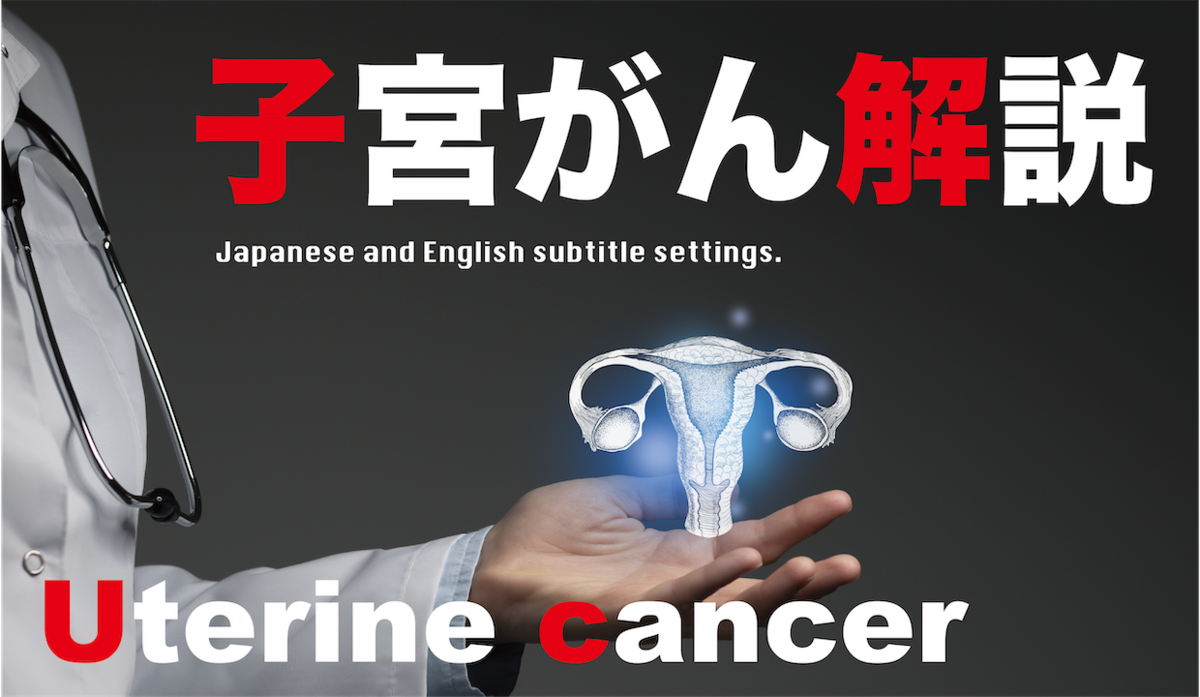
- 子宮がんとは
- 子宮がんの症状
- 子宮がんの発生原因とは
- 子宮がんの治療方法
- 子宮がんの手術後に出やすい合併症
- 子宮がんのステージと生存率
- 子宮がんの予防方法
- 子宮がん患者の体験談
- まとめ
- What is uterine cancer?
- Symptoms of uterine cancer
- What causes uterine cancer?
- How to treat uterine cancer
- Complications that are likely to occur after surgery for uterine cancer
- Urination trouble
- Stage and survival rate of uterine cancer
- How to prevent uterine cancer
- Experiences of uterine cancer patients
- summary
子宮がんは、日本全国で1年間に約1万人以上が診断されており、20歳代後半から患者数が増加しているがんです。
以前は40~50歳代が発症のピークでしたが、最近では20から30歳代の若い女性の発症率が高くなっており、患者数と死亡率は共に増加しています。
今回は、早期発見が重要と言われている子宮がんについて解説していきたいと思います。
子宮がんとは
子宮がんとは、子宮に発生する悪性腫瘍です。
子宮がんは、子宮下部にある筒状の子宮頸部に出来る「子宮頸がん」と、子宮上部の袋状の子宮体部に出来る「子宮体がん」に分かれています。
子宮頸がんは、子宮の入り口付近に発生する事が多く、子宮がんの中では発見されやすいと言われています。
子宮体がんは、子宮の内膜から発生するので、子宮頸がんに比べると発見しづらい種類になります。
子宮がんの症状
では次に、子宮がんの症状について解説していきましょう。
子宮がんは、早期の段階では初期症状はほとんど自覚できないがんでもあります。
しかし、その中でもある程度進行してくと自覚出来る症状が出てきます。
子宮頸がんと子宮体がんでは、症状の現れ方が異なりますので分けて説明していきたいと思います。
子宮頸がんの症状としては、「異常なおりもの」「月経以外の出血」「性行為の際の出血」「下腹部痛」などがあります。
また、濃い茶色の膿「うみ」のようなおりものや、水っぽいおりもの、粘液のようなおりものなど、おりものの異常が分かりやすい症状でしょう。
そして、子宮体がんの一番多い症状は「出血」です。
特に、閉経後に出血がある場合は注意が必要です。
また、「性交時の痛み」「下腹部痛」「腹部膨満感」なども、子宮体がんの症状として出てくる事があります。
子宮がんの発生原因とは
それでは次に、子宮がんの発生原因にはどのようなものがあるのかを見ていきましょう。
子宮がんの中でも「子宮頸がん」と「子宮体がん」では、発生原因が異なります。
まず、子宮頸がんの発生原因から見ていきましょう。
子宮頸がんのほとんどは、ヒトパピローマウイルス(HPV)の感染が原因と言われています。
ヒトパピローマウイルス(HPV)は、性的接触によって感染します。
90%の人は、免疫によってヒトパピローマウイルス(HPV)を排除することが出来ますが、10%の人は排除できず感染が持続するとされているのです。
そして、ヒトパピローマウイルス(HPV)の一部が、がんになる前段階である「前がん病変」を経て、悪性の子宮頸がんへと進行します。
では、子宮体がんの発生原因には、どのようなものがあるのでしょうか。
子宮体がんは、女性ホルモンであるエストロゲンの刺激が長期間続くことで発生する場合と、エストロゲンとは関係ない原因で発生する場合の2通りのパターンがあります。
女性ホルモンであるエストロゲンが関係している場合の原因としては、「出産経験がない」「閉経が遅い」「肥満体型」などが挙げられます。
また、乳がんの治療で使われるタモキシフェンや、更年期障害の治療で使われるエストロゲンを補充する薬を単独で使用する事も、子宮体がんの原因に結びつくと言われています。
そして、エストロゲンとは関係ない原因には、「糖尿病」「血縁者に子宮体がんになった人がいる」というものが挙げられます。
子宮がんの治療方法
それでは次に、子宮がんの治療方法をご紹介していきましょう。
ここでも、子宮頸がんと子宮体がんに分けて、治療方法をご紹介していきたいと思います。
まず子宮頸がんの治療方法には、「手術」「放射線治療」「薬物療法」があります。
単独で行う場合もありますが、それぞれの治療方法を組み合わせて行う事もあります。
手術には、「円錐切除術(えんすいせつじょじゅつ)」「単純子宮摘出術」「準広汎(じゅんこうはん)子宮全摘出術」「広汎(こうはん)子宮全摘出術」「広汎(こうはん)子宮頸部摘出術」があります。
子宮頸部に出来たがんの形や広がり方によって、どのような形で切除するか手術方法を選択します。
放射線治療は、がんに対する主な治療方法の1つで、手術や薬物療法と並んで行われる事もあります。
細胞内のDNAを、高エネルギーのX線やガンマ線などの放射線で直接傷つけることで、治療していく方法です。
ステージに関わらず行う事が出来る治療方法ですが、比較的進行している場合は、細胞障害性抗がん薬とともに放射線治療を行う事が多いです。
そして薬物療法は、主に遠隔転移のある進行がんや、子宮がんが再発した場合に行われる治療方法です。
薬物療法には、「細胞障害性抗がん薬治療」と「分子標的薬治療」があります。
細胞障害性抗がん薬治療では、細胞の増殖の仕組みを利用して、その一部を邪魔する事でがん細胞を攻撃します。
分子標的薬治療は、がん細胞の増殖に関わるタンパク質を標的にして、がんを攻撃する治療方法で細胞障害性抗がん薬とともに使われる事が多いです。
子宮がんの手術後に出やすい合併症
近年では、手術の技術が向上し、手術後の合併症の割合は少なくなってきていますが、一定数の割合では現在でも合併症が起こる事はあります。
ここでは、子宮がんの手術後に起こりやすい合併症についてご紹介していきたいと思います。
リンパ浮腫
子宮がんの手術の際に、リンパ液の通り道であるリンパ節とリンパ管を切り取ることで、リンパ液の通り道が少なくなり足や下腹部がむくみやすくなります。
排尿トラブル
排尿トラブルは、基じん帯を広く切除する「広汎子宮全摘出術」をした場合に起こりやすい合併症と言われています。
「尿が出しにくい」「尿が全部出切らない」「尿がたまった感じがわかりにくい」等の症状があります。
排尿トラブルには個人差がありますが、多くは手術後数週間から数か月程度で回復します。
便秘
便秘は、排尿トラブルと同様に「広汎子宮全摘出術」をした場合に多い合併症です。
比較的、短期間で回復し、排尿トラブルよりも頻度が少ないものになります。
卵巣欠落症状
卵巣欠落症状は、閉経前に両側の卵巣を切除する手術や、放射線治療で卵巣の機能が失われた場合に、女性ホルモンが減少することで起こりやすい合併症です。
具体的には、「ほてり」「発汗」「食欲低下」「イライラ」「頭痛」「肩こり」など、更年期障害のような症状が出てきます。
子宮がんのステージと生存率
子宮がんのステージごとの5年生存率を見ていきましょう。
・ステージ1:93.7%
・ステージ2:77.2%
・ステージ3:57.1%
・ステージ4:21.2%
生存率はあくまで目安です。たとえ進行したステージであっても気をしっかり保つことが重要となります。
子宮がんの予防方法
では、子宮がんの予防方法をご紹介していきたいと思います。
まず、子宮頸がんの予防方法として有効なのは、定期的な検診とワクチンの接種です。
20歳代前半までの女性で、性交渉を経験してから3年以上経っている人の場合は、細胞診という検査方法が有効です。
細胞診は、がんを疑うような異常な細胞がないか調べる検査です。
また、20歳代後半以上の女性の場合は、細胞診とHPV検査が有効です。
HPV検査は、子宮頸がんの原因ウイルスに感染していないかを検査するものです。
そして、子宮体がんの予防方法としては、普段の食事や生活習慣が大きく影響していると言われています。
揚げ物やインスタントラーメンなどを食べる機会が多い人は、子宮体がんの発症リスクが高く、逆に日常的に野菜や豆類を多く食べる人は、子宮体がんの発症リスクが低くなるとされています。
また、子宮体がんは「糖尿病」「肥満体型」なども発症要因とされている為、適度な運動習慣も大切です。
そして、子宮体がんを薬で予防する方法として、低用量ピルが挙げられます。
低用量ピルには、エストロゲンとプロゲステロンの両方が入っているので、ホルモンバランスを整える事ができ、子宮体がんの発症リスクを低下させることが出来るのです。
子宮がん患者の体験談
それでは最後に、子宮がんの患者さん達の体験談をご紹介していきましょう。
37歳 女性
「不正出血がひどい日々が続いて、貧血が気になり婦人科を受診しました。
検査の結果、大きな病院を紹介され翌月には子宮がんと診断されました。
手術で子宮全摘とリンパ節の摘出をし、今は抗がん剤の術後維持療法を開始しています。
抜けてしまった髪も生えてきて、経過観察中です。」
33歳 女性
「20代で子宮がんが見つかり、色々な検査の結果抗がん剤の治療から開始しました。
その後、手術で子宮を全摘し、1週間に1度の抗がん剤は継続しています。
治療開始から吐き気があり、安定剤をもらいながら乗り越えました。
再発の怖さはありますが、前向きに後悔のないように生きていこうと思っています。」
まとめ
今回は、若い女性の発症率が高い、子宮がんについて解説してきました。
子宮がんは、「子宮頸がん」と「子宮体がん」によって予防法や治療方法が異なります。
まずは早期発見が何よりも大切なので、定期的な検診をおすすめします。
===================================================
Uterine cancer is a cancer in which more than 10,000 people are diagnosed every year in Japan, and the number of patients has been increasing since the late 20s.
In the past, the peak onset was in the 40s and 50s, but recently the incidence is higher in young women in the 20s and 30s, and both the number of cases and the mortality rate are increasing.
This time, I would like to explain about uterine cancer, which is said to be important for early detection.
What is uterine cancer?
Uterine cancer is a malignant tumor that begins in the uterus.
Uterine cancer is divided into "cervical cancer" that forms in the tubular cervix in the lower part of the uterus and "cervical cancer" that forms in the sac-like cervix in the upper part of the uterus.
Cervical cancer often occurs near the entrance of the uterus and is said to be more likely to be detected in uterine cancer.
Endometrial cancer originates from the endometrium of the uterus and is a more difficult type to detect than cervical cancer.
Symptoms of uterine cancer
Next, let's explain the symptoms of uterine cancer.
Uterine cancer is also a cancer in which the initial symptoms are hardly noticeable at an early stage.
However, even among them, symptoms that can be noticed appear when it progresses to some extent.
The appearance of symptoms differs between cervical cancer and endometrial cancer, so I would like to explain them separately.
Symptoms of cervical cancer include "abnormal vaginal discharge," "bleeding other than menstruation," "bleeding during sexual activity," and "lower abdominal pain."
In addition, abnormalities in vaginal discharge such as dark brown pus "Umi", watery vaginal discharge, and mucus-like vaginal discharge are easy to understand.
And the most common symptom of endometrial cancer is "bleeding".
Be especially careful if you have bleeding after menopause.
In addition, "pain during sexual intercourse," "lower abdominal pain," and "abdominal bloating" may also appear as symptoms of endometrial cancer.
What causes uterine cancer?
Next, let's see what are the causes of uterine cancer.
Among uterine cancers, "cervical cancer" and "endometrial cancer" have different causes.
First, let's look at the causes of cervical cancer.
Most cervical cancers are said to be caused by infection with the human papillomavirus (HPV).
Human papillomavirus (HPV) is transmitted by sexual contact.
It is said that 90% of people can eliminate human papillomavirus (HPV) by immunity, but 10% cannot eliminate it and the infection persists.
Then, a part of human papillomavirus (HPV) progresses to malignant cervical cancer through "precancerous lesion" which is the stage before cancer.
So what are the causes of endometrial cancer?
There are two patterns of endometrial cancer, one is caused by long-term stimulation of the female hormone estrogen, and the other is caused by a cause unrelated to estrogen.
Causes related to the female hormone estrogen include "no experience of childbirth," "late menopause," and "obese body type."
In addition, it is said that the use of tamoxifen used in the treatment of breast cancer and the drug that supplements estrogen used in the treatment of menopause alone is also linked to the cause of endometrial cancer.
And the causes that are not related to estrogen include "diabetes" and "some relatives have endometrial cancer".
How to treat uterine cancer
Next, let's introduce how to treat uterine cancer.
Here, too, I would like to introduce treatment methods separately for cervical cancer and endometrial cancer.
First of all, there are "surgery", "radiotherapy" and "drug therapy" as treatment methods for cervical cancer.
It may be given alone, or it may be given in combination with each treatment method.
Surgery includes "conization", "simple hysterectomy", "quasi-extensive hysterectomy", "extensive hysterectomy", and "extensive hysterectomy". ) There is a "cervical conization".
Depending on the shape and spread of the cancer in the cervix, the surgical method should be selected.
Radiation therapy is one of the main treatments for cancer and may be given alongside surgery or medication.
It is a method of treating the intracellular DNA by directly damaging it with high-energy X-rays, gamma rays, and other radiation.
It is a treatment method that can be performed regardless of the stage, but if it is relatively advanced, radiation therapy is often performed together with a cytotoxic anticancer drug.
And drug therapy is a treatment method that is mainly used when advanced cancer with distant metastasis or uterine cancer recurs.
Pharmacotherapy includes "cytotoxic anticancer drug treatment" and "molecular-targeted drug treatment".
In cytotoxic anticancer drug treatment, cancer cells are attacked by using the mechanism of cell proliferation and interfering with some of them.
Molecular-targeted drug therapy is a treatment method that targets proteins involved in the growth of cancer cells and attacks cancer, and is often used together with cytotoxic anticancer drugs.
Complications that are likely to occur after surgery for uterine cancer
In recent years, surgical techniques have improved and the rate of postoperative complications has decreased, but complications may still occur at a certain rate.
Here, I would like to introduce the complications that are likely to occur after surgery for uterine cancer.
Lymphedema
By cutting out the lymph nodes and lymph vessels that are the paths of lymph during surgery for uterine cancer, the lymph nodes are reduced and the legs and lower abdomen are more likely to swell.
Urination trouble
Urination problems are said to be a complication that tends to occur when performing "extensive hysterectomy", which removes a wide area of the ligament.
There are symptoms such as "difficulty in urinating", "not enough urine", and "difficulty in understanding the feeling of urine accumulation".
There are individual differences in urination problems, but most of them recover within a few weeks to a few months after surgery.
constipation
Constipation is a common complication of "extensive hysterectomy" as well as urination problems.
It recovers in a relatively short period of time and is less frequent than urination problems.
Ovarian deficiency symptoms
Ovarian deficiency symptoms are a common complication of decreased female hormones when surgery to remove the ovaries on both sides before menopause or when radiation therapy causes the ovaries to lose their function.
Specifically, symptoms such as menopause appear, such as "hot flashes," "sweat," "decreased appetite," "irritability," "headache," and "stiff shoulders."
Stage and survival rate of uterine cancer
Let's look at the 5-year survival rate for each stage of uterine cancer.
・ Stage 1: 93.7%
・ Stage 2: 77.2%
・ Stage 3: 57.1%
・ Stage 4: 21.2%
Survival rate is just a guide. It's important to stay alert, even if it's an advanced stage.
How to prevent uterine cancer
Now, I would like to introduce how to prevent uterine cancer.
First, regular screening and vaccination are effective ways to prevent cervical cancer.
For women in their early twenties who have had sexual intercourse for more than three years, a test called cytology is effective.
Cytology is a test to check for abnormal cells that may be suspected of having cancer.
For women in their late 20s and older, cytology and HPV testing are effective.
The HPV test tests for infection with the virus that causes cervical cancer.
And, as a preventive method for endometrial cancer, it is said that the usual diet and lifestyle have a great influence.
People who often eat fried foods and instant noodles have a high risk of developing endometrial cancer, while those who eat a lot of vegetables and legumes on a daily basis have a low risk of developing endometrial cancer. ..
In addition, since endometrial cancer is also considered to be a cause of onset such as "diabetes" and "obese body type", proper exercise habits are also important.
And low-dose pills are a way to prevent endometrial cancer with drugs.
Low-dose pills contain both estrogen and progesterone, which can help balance the hormones and reduce the risk of developing endometrial cancer.
Experiences of uterine cancer patients
Finally, let me introduce the experiences of patients with uterine cancer.
37 year old female
"I went to the gynecology department because I was worried about anemia because of the terrible days of irregular bleeding.
As a result of her examination, she was referred to a large hospital and was diagnosed with uterine cancer the following month.
She had surgery for total hysterectomy and lymph node removal, and is now starting postoperative maintenance therapy with anticancer drugs.
Her hair that has fallen out is also growing and is being followed up. "
33 year old female
"Uterine cancer was found in my twenties, and as a result of various tests, I started treatment with anticancer drugs.
After that, she had her uterus removed by surgery, and she continues to take anticancer drugs once a week.
She had nausea from the start of treatment and overcame it with a stabilizer.
I am afraid of recurrence, but I will live positively and without regrets. "
summary
This time, I have explained about uterine cancer, which has a high incidence in young women.
For uterine cancer, prevention and treatment methods differ depending on whether it is "cervical cancer" or "endometrial cancer."
First of all, early detection is the most important, so regular examinations are recommended.
乳がんについて About Breast cancer.
*There is an English translation in the second half of the article.
- 乳がんとは
- 乳がんの症状
- 乳がんの原因
- 乳がんの治療方法
- 乳がんの病期(ステージ)
- 乳がんの生存率
- 乳がんの治療にかかる費用
- 乳がんの検診方法
- 乳がんのセルフチェック方法
- 乳がんの患者さんの体験談
- まとめ
- What is breast cancer?
- Breast cancer symptoms
- Causes of breast cancer
- How to treat breast cancer
- Breast cancer stage
- Breast cancer survival rate
- The cost of treating breast cancer
- Breast cancer screening method
- Breast cancer self-check method
- Breast Cancer Patients' Experiences
- summary
乳がんは、日本人の女性がかかるがんとしては、最も頻度が高いがんと言われています。
日本全国で1年間に、およそ92,300人が乳がんと診断されています。
全体では女性の発症率が高いですが、男性でも1年間に約650人が乳がんと診断されているのです。
40歳代後半~60歳代後半の発症率が高いですが、若い人が乳がんになると、転移するスピードも速いため早期発見が重要ながんでもあります。
今回は、そんな乳がんについて詳しく解説していきたいと思います。
乳がんとは
乳がんとは、乳腺の組織にがんが発生する病気です。
多くの場合は、乳管から発生し、女性だけでなく男性にも発生するがんでもあります。
転移の可能性が非常に高いがんで、乳房のまわりのリンパ節や、離れた臓器へ転移する事も多いのです。
乳がんは、主に「非浸潤がん」と「浸潤がん」に分ける事が出来ます。
非浸潤がんは、乳管や乳腺小葉にがん細胞がとどまっているがんを指し、浸潤がんは乳管や乳腺小葉のまわりにも広がるがんです。
またその他にも、特殊な乳がんとして「粘液がん」「専用の腺様嚢胞(せんようのうほう)がん」などの炎症性の乳がんなどもあります。
乳がんの症状
それでは次に、乳がんの症状を解説していきましょう。
乳がんの症状で一番多いのが、「しこり」です。
乳房や乳頭の裏などに現われる事が多いですが、しこり自体は乳房周辺さまざまな所に出来ます。
しこりは、乳がんが進行するにつれ大きくなり、早期での発見は難しいのが現状です。
そして、「乳房の左右差」や「えくぼのような凹み」「ただれ」や「乳頭からの出血」「リンパ節の腫れ」なども乳がんの症状として有名です。
乳房の左右差は、乳がんでなくても見られることもありますが、以前までは左右対称だったのに大きさや硬さが変わった場合は注意が必要です。
えくぼのような凹みは、乳がんで出来たしこりによって皮膚や乳頭が引っ張られる事で起こります。
そのため、しこり自体が発見できなくても、皮膚の引き連れが症状のサインとして現れる事もあるのです。
乳頭や乳輪のまわりがただれたり、かさぶたが出来るのは、パジェット病という乳がんの症状です。
さらに、乳がんは母乳が通る管である乳管に出来ることも多く、その部分からの出血も乳がんの症状として多いでしょう。
リンパ節の腫れは、乳がんが転移している時に現われる症状として多く、腕がむくんだり、わきの下にしこりが現れたりします。
乳がんの原因
乳がんの発生原因として考えられているのは、女性ホルモンである「エストロゲン」が大きく関わっているという事です。
閉経後のホルモン補充療法や、経口避妊薬などの使用は、乳がんを発生させるリスクが高いとも言われているのです。
また乳がんは、生活習慣とも密接に関わっており、閉経後の肥満、飲酒、運動不足なども発生リスクを高める要因となります。
そして、初経年齢が低かったり、出産経験がない、授乳経験がないなどの他に、異型乳管過形成をはじめとした異型を伴う上皮内病変にかかった経験がある場合も、乳がんの発生リスクを高めるようです。
さらに、乳がんは遺伝性が高い病気でもあります。
第一親等で乳がんになった血縁者がいる場合も、発生リスクは高まるとされています。
乳がんの治療方法
では次に、乳がんの治療方法について解説していきたいと思います。
乳がんの治療には、大きく分けて「手術」「放射線治療」「薬物療法」の3種類があります。
手術を行う場合は、「乳房部分切除術」と「乳房全切除術」のどちらかを行うのが一般的です。
乳房部分切除術は、手術後に放射線照射を行い再発を防ぐ事を前提として行われ、がんを確実に取り除きながら、患者さんが美容的に満足できる乳房を残すことを目的として行われます。
乳房全切除術は、乳がんが広範囲に広がっている場合に行われる手術方法です。
放射線治療は、がんに直接高エネルギーのX線を照射して、がん細胞を死滅させたり小さくさせる治療法です。
通常1日1回、週5回の照射を約4~6週間かけて行います。
薬物療法は、「ホルモン療法薬」「分子標的薬」「細胞障害性抗がん薬」などを用いて行う治療法です。
薬物療法は、再発の危険性を下げたり、症状を緩和する目的で行われることが多く、病期やリスクに応じて行われます。
乳がんの病期(ステージ)
それでは、乳がんの進行程度ごとの病期(ステージ)をご紹介していきます。
・0期:非浸潤がんまたは、パジェット病による早期のがん
・Ⅰ期:がんの大きさが2cm以下で、リンパ節やまわりの臓器への転移がない状態
・ⅡA期:がんの大きさが2cm以下で、わきの下のリンパ節に転移があるが固定されておらず動く状態、もしくはがんが2cm以上5cm以下の大きさでリンパ節やまわりの臓器への転移がない状態。
・ⅡB期:がんが2cm以上5cm以下で、わきの下のリンパ節に転移があるが固定されておらず動く状態、もしくはがんが5cm以上でリンパ節やまわりの臓器への転移がない状態。
・ⅢA期:がんが5cm以下で、わきの下のリンパ節に転移していて固定されているがリンパ節が互いに癒着している状態、またはわきの下のリンパ節には転移していないが胸骨の内側のリンパ節に転移している状態、もしくはしこりが5cm以上でわきの下または胸骨の内側のリンパ節に転移がある状態。
・ⅢB期:がんの大きさやリンパ節への転移に関わらず、しこりが胸壁に固定されていたり、がんが皮膚に出たり崩れたりむくんで見える状態、もしくはしこりはないが炎症性乳がんなどもこの病期に含まれる。
・ⅢC期:がんの大きさに関わらず、わきの下のリンパ節と胸骨の内側のリンパ節の両方に転移がある状態、もしくは鎖骨の上下にあるリンパ節に転移がある状態。
・Ⅳ期:他の離れた臓器(骨・肺・肝臓・脳など)への転移がある状態。
乳がんの生存率
乳がんの生存率は、ステージによって変わってきます。
早期に発見出来れば、治癒する可能性も高く、治療法によっても異なります。
ここでご紹介する数値は、あくまで平均的な目安として参考にしてみてください。
・ステージ0:約98%
・ステージ1:約98%
・ステージ2:約87%
・ステージ3:約77%
・ステージ4:約20%
乳がんの治療にかかる費用
乳がんの治療にかかる費用は、治療法や期間などでも変わってきますが、ここでは平均的な費用をご紹介していきたいと思います。
一般的に乳がんの手術のみで、乳房部分切除術が30~40万円、乳房全切除術が20~50万円となっています。
この他に、検査費用・入院費用・その他の治療費用を含めて、健康保険適応(3割負担)で約20~40万円前後となっています。
放射線治療に関しては、1年目で約33万円、抗がん剤費用では約60~70万円ほどとなっています。
2年目の再発予防抗がん剤治療の費用が、130~140万円程度となっています。
乳がんの検診方法
それでは、乳がんを発見する為に重要な、検診の流れをご紹介していきましょう。
- 問診:月経周期や出産歴など、気になる症状の有無などの問診を行います。
- 視触診検査:医師が乳房のしこりや変形などがないか、視触診でチェックします。
- 画像検査:マンモグラフィーや超音波検査で、画像化した状態で乳房内部をチェックします。
- 診断結果:医師によるチェックで、乳がんの可能性の診断結果を受け取ります。
このような流れで、乳がんの検診が行われます。
日頃からセルフチェックをしていると、問診の際に気になる事を聞きやすくなるので、セルフチェックをすることもおすすめします。
乳がんのセルフチェック方法
では次に、乳がんを早期発見する為のセルフチェック方法をご紹介していきましょう。
乳がんを、初期に発見する為には、一番気づきやすい「しこり」をセルフチェックで見つける事が重要です。
しこりを見つける為には、手のひらを使って乳房を全体的に触ります。
乳頭の裏や、乳房の外側にしこりは出来やすいため、少し力を入れて奥まで触るようにしましょう。
この時に、固くて動きにくく、境界がはっきりしているしこりが手に触れた場合は、医療機関を受診する事をおすすめします。
また、日頃から乳房の形や向きなどを、定期的に観察する事もセルフチェックでは欠かせません。
毎日見ているからこそ気づく変化があるからです。
皮膚の引き連れや、乳頭にくぼみがないかなど、チェックするようにしましょう。
乳がんの患者さんの体験談
それでは最後に、乳がんの患者さんの体験談をご紹介していきたいと思います。
・50代女性
「人間ドックのマンモグラフィーで、しこりが発見されました。健診結果が届くまでの2週間は心が壊れそうでした。
ステージは3で悪性度は4、腫瘍の大きさは3cmほどでした。ホルモン薬を使っての治療でしたが、食欲やメンタル面の維持が大変でした。
退院後は、鉛筆で線がまっすぐ書けないなど変わった部分もありますが、家族や友人などまわりの人達に支えられて、再発予防をしながら生活しています。」
・30代女性
「私は34歳の時に、突然「若年性トリプルネガティブ乳がん」になりました。
検査後1ヶ月後に手術を受けましたが、がんの勢いは驚くほど速くリンパ節転移アリの状態でした。
再発率の高い乳がんで、手術後は抗がん剤治療を8クールと放射線25回の治療を行いました。今は、再発予防を徹底して、家庭と治療・仕事の両立に頑張っています。」
まとめ
さて今回は、乳がんについて解説してきました。
乳がんは、がんの中でも早期発見が難しいと言われ、その分セルフチェックや検診が重要になってきます。
ステージによって治療法や生存率も変わるので、いかに早く発見するかがカギとなるのです。
================================
Breast cancer is said to be the most common cancer that affects Japanese women.
Approximately 92,300 people are diagnosed with breast cancer in a year across Japan.
Overall, the incidence is high in females, but about 650 males are diagnosed with breast cancer each year.
The incidence is high in the late 40s to late 60s, but when young people develop breast cancer, the speed of metastasis is fast, so early detection is also important.
This time, I would like to explain in detail about such breast cancer.
What is breast cancer?
Breast cancer is a disease in which cancer develops in the tissues of the mammary glands.
Often, it is a cancer that begins in the breast ducts and affects men as well as women.
Cancer with a very high probability of metastasis often spreads to the lymph nodes around the breast and to distant organs.
Breast cancer can be divided into "non-invasive cancer" and "invasive cancer".
Non-invasive cancer refers to cancer in which cancer cells remain in the ducts and lobules of the breast, and invasive cancer is cancer that spreads around the ducts and lobules of the breast.
In addition, there are also inflammatory breast cancers such as "mucinous cancer" and "dedicated adenoid cystic carcinoma" as special breast cancers.
Breast cancer symptoms
Next, let's explain the symptoms of breast cancer.
The most common symptom of breast cancer is "lumps".
It often appears on the back of the breast or nipple, but the lump itself can be found in various places around the breast.
The lump grows larger as breast cancer progresses, making it difficult to detect at an early stage.
Breast cancer symptoms such as "left-right difference in breast", "dimple-like dent", "swelling", "bleeding from nipple", and "swelling of lymph nodes" are also famous.
Left-right differences in the breast can be seen even if you don't have breast cancer, but you should be careful if the size or hardness changes, even though it was previously symmetrical.
Dimple-like dents are caused by the lumps created by breast cancer pulling on the skin and nipples.
Therefore, even if the lump itself cannot be found, skin accompaniment may appear as a sign of symptoms.
Sores around the nipples and areola and scabs are symptoms of Paget's disease, a breast cancer.
In addition, breast cancer often forms in the ducts through which breast milk passes, and bleeding from that area is also a common symptom of breast cancer.
Swelling of the lymph nodes is a common symptom of breast cancer metastasis, with swelling of the arms and lumps under the armpits.
Causes of breast cancer
The cause of breast cancer is thought to be that the female hormone "estrogen" is greatly involved.
Postmenopausal hormone replacement therapy and the use of oral contraceptives are also said to have a high risk of developing breast cancer.
Breast cancer is also closely related to lifestyle-related habits, and postmenopausal obesity, alcohol consumption, and lack of exercise are also factors that increase the risk of developing breast cancer.
And if you have a younger age, have no experience of childbirth, have no breastfeeding experience, or have had atypical intraepithelial lesions such as atypical ductal hyperplasia, you are also at risk of developing breast cancer. Seems to increase.
In addition, breast cancer is a highly hereditary disease.
It is also said that the risk of developing breast cancer is increased if there are relatives who have breast cancer in the first degree of kinship.
How to treat breast cancer
Next, I would like to explain how to treat breast cancer.
There are three main types of breast cancer treatment: surgery, radiation therapy, and drug therapy.
When performing surgery, it is common to perform either "partial mastectomy" or "total mastectomy".
Partial breast resection is performed on the premise that radiation is given after surgery to prevent recurrence, and the purpose is to leave a breast that is cosmetically satisfying for the patient while reliably removing the cancer.
Total mastectomy is a surgical procedure performed when breast cancer is widespread.
Radiation therapy is a treatment that kills or shrinks cancer cells by directly irradiating the cancer with high-energy X-rays.
Irradiation is usually performed once a day, 5 times a week for about 4 to 6 weeks.
Pharmacotherapy is a treatment method that uses "hormone therapy drugs," "molecular-targeted drugs," "cytotoxic anticancer drugs," etc.
Drug therapy is often given to reduce the risk of recurrence or alleviate symptoms, depending on the stage and risk.
Breast cancer stage
Then, I will introduce the stage of breast cancer according to the degree of progression.
・ Stage 0: Non-invasive cancer or early stage cancer due to Paget's disease
・ Stage I: The size of the cancer is 2 cm or less, and there is no metastasis to lymph nodes or surrounding organs.
・ Stage IIA: Cancer is 2 cm or less in size and has metastases to the lymph nodes under the arm but is not fixed and moves, or cancer is 2 cm or more and 5 cm or less in size to the lymph nodes and surrounding organs. No metastasis.
・ Stage IIB: Cancer is 2 cm or more and 5 cm or less, and there is metastasis to the lymph nodes under the arm, but it is not fixed and moves, or cancer is 5 cm or more and there is no metastasis to the lymph nodes or surrounding organs.
Stage IIIA: Cancer is 5 cm or less and has spread to and fixed to the lymph nodes under the arm, but the lymph nodes are attached to each other, or have not spread to the lymph nodes under the arm but are inside the thoracic bone. Metastases to the lymph nodes in the area, or metastases to the lymph nodes under the armpit or inside the thoracic bone with a lump of 5 cm or more.
・ Stage IIIB: Regardless of the size of the cancer or metastasis to lymph nodes, lumps are fixed to the chest wall, the cancer appears on the skin, collapses, or appears swollen, or there is no lump but inflammatory breast cancer. Is also included in this stage.
Stage IIIC: Metastases to both the lymph nodes under the arm and the lymph nodes inside the sternum, or to the lymph nodes above and below the clavicle, regardless of the size of the cancer.
・ Stage IV: Metastasis to other distant organs (bone, lung, liver, brain, etc.).
Breast cancer survival rate
Breast cancer survival varies from stage to stage.
If it can be detected early, it is likely to be cured and it depends on the treatment method.
Please refer to the numbers introduced here as an average guide only.
・ Stage 0: Approximately 98%
・ Stage 1: Approximately 98%
・ Stage 2: Approximately 87%
・ Stage 3: Approximately 77%
・ Stage 4: Approximately 20%
The cost of treating breast cancer
The cost of treating breast cancer varies depending on the treatment method and duration, but here I would like to introduce the average cost.
Generally, only breast cancer surgery costs 300,000 to 400,000 yen for partial breast resection and 200,000 to 500,000 yen for total breast resection.
In addition to this, including examination costs, hospitalization costs, and other treatment costs, the health insurance coverage (30% burden) is about 200,000 to 400,000 yen.
Radiation therapy costs about 330,000 yen in the first year, and the cost of anticancer drugs is about 600,000 to 700,000 yen.
The cost of anti-cancer drug treatment to prevent recurrence in the second year is about 1.3 to 1.4 million yen.
Breast cancer screening method
Now, let's introduce the flow of screening, which is important for detecting breast cancer.
Interview: Interviews will be conducted regarding the presence or absence of symptoms of concern such as the menstrual cycle and childbirth history.
Palpation: Your doctor will check your breasts for lumps or deformities by palpation.
Imaging: A mammography or ultrasonography is used to check the inside of the breast in an imaged state.
Diagnosis Results: Checked by your doctor, you will receive a diagnosis of possible breast cancer.
Breast cancer screening is carried out in this way.
If you do a self-check on a regular basis, it will be easier to hear what you are worried about when interviewing, so it is also recommended to do a self-check.
Breast cancer self-check method
Next, let's introduce a self-check method for early detection of breast cancer.
In order to detect breast cancer in the early stages, it is important to find the most noticeable "lump" by self-check.
To find the lump, use your palm to touch the entire breast.
It's easy to get lumps on the back of the nipple or on the outside of the breast, so use a little force and touch it all the way.
At this time, if you feel a lump that is hard, difficult to move, and has a clear boundary, it is recommended that you consult a medical institution.
In addition, it is indispensable for self-check to regularly observe the shape and orientation of the breast.
This is because there are changes that you will notice because you are watching every day.
Make sure to check for skin dents and nipple dents.
Breast Cancer Patients' Experiences
Finally, I would like to introduce the experiences of breast cancer patients.
・ Female in her 50s
"A lump was found on the mammography of the human dock. It was almost heartbreaking for two weeks before the results of the medical examination arrived.
The stage was 3, the malignancy was 4, and the size of the tumor was about 3 cm. I was treated with hormonal drugs, but it was difficult to maintain my appetite and mental health.
After I was discharged from the hospital, there were some strange things such as not being able to draw straight lines with a pencil, but I am living while preventing recurrence with the support of people around me such as my family and friends. "
・ Female in her 30s
"When I was 34, I suddenly had" juvenile triple-negative breast cancer. "
I had surgery one month after the test, but the cancer was surprisingly fast and was in the state of lymph node metastases.
For breast cancer with a high recurrence rate, after surgery, 8 courses of anticancer drug treatment and 25 times of radiation treatment were performed. Now, I am working hard to prevent recurrence and to balance treatment and work with my family. "
summary
Well, this time, I have explained about breast cancer.
Breast cancer is said to be difficult to detect at an early stage among cancers, and self-checks and screenings are important accordingly.
Treatment and survival rates vary depending on the stage, so the key is how quickly you can detect them.
大腸がんについて About Colorectal cancer

*There is an English translation in the second half of the article.
- 大腸がんとは
- 大腸がんの症状
- 大腸がんの患者数
- 大腸がんの原因とは
- 大腸がんの検査や診断
- 大腸がんの治療や費用
- 大腸がんの生存率
- 大腸がん患者の体験談
- まとめ
- What is colorectal cancer?
- Colorectal cancer symptoms
- Number of patients with colorectal cancer
- What Causes Colorectal Cancer?
- Colorectal cancer testing and diagnosis
- Treatment and costs of colorectal cancer
- Colorectal cancer survival rate
- Experiences of colorectal cancer patients
- summary
大腸がんは、日本で発症率の高いがんの中の1つで、毎年約8万人前後が発症していると言われています。
しかし、化学療法の進歩によって、治療成績の向上が著しい病気でもあり、早期発見出来れば予後の良いがんでもあります。
今回は、そんな大腸がんについて詳しく解説していきたいと思います。
大腸がんとは
まず、大腸がんとはどのような病気なのか解説していきましょう。
大腸がんは、大腸の粘膜から発生する悪性腫瘍の1つです。
がんが出来る場所によって、「結腸がん」と「直腸がん」の2種類に分けられます。
大腸は、入り口から出口に向かうまでのいくつかの部位に分かれていて、「盲腸」「上行結腸」「横行結腸」「下行結腸」「S状結腸」「直腸S字部」「上部直腸」「下部直腸」「肛門菅」で構成されています。
この中で、大腸がんは特に「S状結腸」や「直腸」に発生する割合が多いと言われているのです。
大腸がんの症状
それでは次に、大腸がんの症状を詳しく解説していきたいと思います。
大腸がんは、初期の段階では自覚症状があまりないのが特徴です。
そのため、自覚症状が出た時には、がんが進行しているケースが多いがんでもあります。
大腸がんが進行すると、「血便」「下血」「下痢や便秘」「体重減少」「継続的な血便や下血による貧血」などの症状が現れてきます。
さらに大腸がんが大きくなってくると、お腹の中でしこりを感じるようになります。
また、がんの影響で腸閉塞を起こし、便が出にくくなったり、腹痛や嘔吐のような症状が出る事もあります。
大腸がんの症状の出方は、がんが発生している部位によっても変わってきます。
大腸の出口である、自分から見て左側に大腸がんの腫瘍がある場合は、早期から先ほどご紹介した症状が出始めます。
しかし、大腸の入り口付近や中心の辺りの腸が太い部分に、がんが発生している場合は、症状が出始めるのが腫瘍がある程度大きくなってからという事も少なくありません。
大腸がんの患者数
日本では大腸がんにかかる患者数は、40代から増加すると言われています。
日本で1年間に新たに大腸がんと診断された患者数は、2017年では男性が約9万人、女性は約7万人となっています。
また、日本人10万人あたりの罹患率を臓器別に見てみても、大腸がんは男性は3番目、女性は2番目に多いがんとなっています。
大腸がんは、女性よりも男性に多い傾向にあり、年齢が上がるにつれて増えていくという特徴があるのです。
大腸がんの原因とは
では、大腸がんの原因とはどのようなものが考えられているのでしょうか。
大腸がんの原因は、私たちのライフスタイルや食事の影響が大きいと考えられています。
・食の欧米化
例えば、食生活の欧米化です。
昔に比べると、現在の日本人の食事は、脂質や動物性たんぱく質の摂取量が多い欧米化になっています。
またその一方で、炭水化物や食物繊維の摂取量が減っているのが現状です。
このような食事スタイルによって、便が大腸内に停滞する時間が長くなり、腸内環境が悪化する原因となるのです。
・運動不足
また、運動不足も大腸がんの原因の1つと考えられています。
自動車の発達によって、日本人の歩数が昔に比べると確実に減少しています。
このことによって、体の中の巡りが悪くなり、発がん物質が大腸粘膜に長く停滞する要因となるのです。
・遺伝性
そして、数は少ないながらも大腸がんの原因として考えられるのが、遺伝性の大腸がんです。
遺伝性で多いのが、「家族性大腸ポリポーシス」と「遺伝性非ポリポーシス大腸がん」です。
「家族性大腸ポリポーシス」は、大腸がん全体の約1%を占めていて、若い年代で大腸の中にポリープが無数に出来て、放置すれば100%がん化します。
「遺伝性非ポリポーシス大腸がん」は、ポリープを伴わない大腸がんで、大腸がん全体の約5%を占めています。
大腸がんの検査や診断
大腸がんの検査は、まず便潜血検査で便に血液が混じっていないかを調べるところから始まります。
もし便潜血検査で陽性になれば、大腸内視鏡検査や注腸造影検査を行い、さらに詳しく調べていきます。
大腸内視鏡検査や注腸造影検査によって、がんが発見された場合は、さらにCT検査、MRI検査によって、腫瘍の大きさや大腸の状態を調べます。
大腸内視鏡検査では、主に腫瘍の大きさや形、色や広がりなどを画像によって確認します。
また注腸造影検査では、がんがどこに出来ているか正確な位置や深さを調べます。
その他に、肛門から直接触ってしこりを確認する直腸指診や、採血での腫瘍マーカー検査、腫瘍の一部を切り取って顕微鏡で調べる病理検査、放射線の薬剤を使って全身への転移を調べるPET検査などがあります。
このような検査を症例に応じて行い、転移の有無や進行度合いなどが最終的に診断されます。
大腸がんの治療や費用
では次に、大腸がんの治療や費用について解説していきたいと思います。
大腸がんの治療は、がんの進行度合いや患者の年齢によって、適切な方法に分けられます。
例えば、比較的早期に大腸がんを発見できた場合は、転移の可能性も低いため内視鏡を使って腫瘍を切除するという治療方法が多いです。
一方、がんが進行して根が深くなっている場合は、他の臓器への転移の可能性が高いため、開腹手術や腹腔鏡手術が行われます。
このように、がんが発生している場所や進行度合いによって治療方法は異なりますが、基本的には腫瘍のある部分とリンパ節を取り、他の臓器や組織の切除なども行います。
腸管を切除した場合は、残っている腸管とつなぎ合わせ、放射線治療や抗がん剤治療を併せて行うのが一般的です。
では次に、大腸がんの検査や治療にかかる費用についてです。
患者さんの医療費負担が3割の場合。
「検査の費用」
・腫瘍マーカー:690円
・大腸内視鏡検査:約10,000円
・注腸造影検査:約4,000円
・造影CT:約9,000円
・造影MRI:約9,000円
・PET-CT:約27,000円
「内視鏡治療の費用」
・ポリペクトミー:約20,000円
・内視鏡的粘膜切除術:約26,000円
・内視鏡的粘膜下層剥離術:約70,000円
「手術にかかる費用」
・結腸がんで手術し約2週間入院した場合:約30万円~40万円
・直腸がんで手術し約2週間入院した場合:約40万円~50万円
大腸がんの生存率
では次に、大腸がんの患者さんの、ステージごとの平均的な生存率をご紹介します。
ここでは、ステージ別の5年生存率を割合で示しています。
・ステージ0:94%
・ステージⅠ:91.6%
・ステージⅡ:84.8%
・ステージⅢ:77.7~60%
・ステージⅣ:18.8%
大腸がん患者の体験談
それでは最後に、大腸がんの患者さんの体験談をご紹介していきましょう。
・65歳 男性
「普段から、体の変化に敏感で大腸がんの診断をされた時も不思議と冷静でした。元来前向きな性格で、そうした自分の性格もがんから生還できた理由かもしれません。
治療方針などは、すべて医師に任せ「痛い」「つらい」という感情は我慢せずに伝えるようにしていました。抗がん剤の副作用は辛いものでしたが、手術して、1年の治療後は精神的にも肉体的にもだいぶ楽になりました。」
・60歳 男性
「旅行先で腸閉塞になり、その翌日も嘔吐でダウンして病院に駆け込みました。診察の結果大腸がんの診断をされ、手術を行う事になりました。
リンパ転移のステージⅢ。怖い気持ちと闘いながら、術後の経過もスッキリしない日々を過ごしました。出来ない事が多くなりましたが、抗がん剤治療を行いながら前向きに治療しています。」
まとめ
さて今回は、日本で発症率の高いがんの1つである「大腸がん」について解説してきました。
大腸がんの原因は様々ですが、日頃の食生活やライフスタイルに気をつける事で、予防できる病気でもあります。
定期的な検査と合わせて、年齢とともに上がる大腸がんのリスクから体を守りましょう。
大腸癌
==============================
Colorectal cancer is one of the cancers with the highest incidence in Japan, and it is said that about 80,000 people develop it every year.
However, it is also a disease whose treatment results are significantly improved due to advances in chemotherapy, and it is also a cancer with a good prognosis if it can be detected early.
This time, I would like to explain in detail about such colorectal cancer.
What is colorectal cancer?
First, let's explain what kind of disease is colorectal cancer.
Colorectal cancer is a malignant tumor that begins in the mucous membrane of the large intestine.
Depending on where the cancer develops, it can be divided into two types: colon cancer and rectal cancer.
The colon is divided into several parts from the entrance to the exit. It consists of the lower rectum and the anal canal.
Of these, colorectal cancer is said to occur most often in the "sigmoid colon" and "rectum."
Colorectal cancer symptoms
Next, I would like to explain the symptoms of colorectal cancer in detail.
Colorectal cancer is characterized by few subjective symptoms in the early stages.
Therefore, when subjective symptoms appear, it is often the case that the cancer has progressed.
As colorectal cancer progresses, symptoms such as "bloody stool", "melena", "diarrhea and constipation", "weight loss", and "anemia due to continuous bloody stool and melena" appear.
As colorectal cancer grows larger, you will feel a lump in your stomach.
In addition, the effects of cancer may cause intestinal obstruction, making it difficult to get stools and causing symptoms such as abdominal pain and vomiting.
The appearance of symptoms of colorectal cancer also depends on where the cancer is occurring.
If you have a colon cancer tumor on the left side of your large intestine, which is the exit of the large intestine, the symptoms mentioned earlier will begin to appear from an early stage.
However, if cancer develops near the entrance to the large intestine or in the thick part of the intestine near the center, it is not uncommon for symptoms to begin to appear after the tumor has grown to some extent.
Number of patients with colorectal cancer
In Japan, the number of patients with colorectal cancer is said to increase from the 40s.
In 2017, the number of patients newly diagnosed with colorectal cancer in Japan was about 90,000 for men and about 70,000 for women.
Looking at the prevalence per 100,000 Japanese by organ, colorectal cancer is the third most common cancer in men and the second most common in women.
Colorectal cancer tends to be more common in men than in women, and is characterized by an increase with age.
What Causes Colorectal Cancer?
So what are the possible causes of colorectal cancer?
The cause of colorectal cancer is thought to be largely influenced by our lifestyle and diet.
・ Westernization of food
For example, the westernization of eating habits.
Compared to the past, the current Japanese diet has become westernized with a high intake of lipids and animal proteins.
On the other hand, the current situation is that the intake of carbohydrates and dietary fiber is decreasing.
This dietary style causes the stool to stay in the large intestine for a longer period of time, causing the intestinal environment to deteriorate.
·Lack of exercise
Lack of exercise is also thought to be one of the causes of colorectal cancer.
With the development of automobiles, the number of steps taken by Japanese people is steadily decreasing compared to the past.
This makes it difficult to circulate in the body and causes carcinogens to stay in the mucous membrane of the large intestine for a long time.
・ Hereditary
And, although the number is small, hereditary colorectal cancer is considered to be the cause of colorectal cancer.
The most hereditary are "familial adenomatous polyposis" and "hereditary nonpolyposis colorectal cancer".
"Familial adenomatous polyposis" accounts for about 1% of all colorectal cancers, and innumerable polyps form in the large intestine at a young age, and if left untreated, it becomes 100% cancerous.
"Hereditary nonpolyposis colorectal cancer" is a colorectal cancer without polyps and accounts for about 5% of all colorectal cancers.
Colorectal cancer testing and diagnosis
A colorectal cancer test begins with a fecal occult blood test to check for blood in the stool.
If the fecal occult blood test is positive, a colonoscopy or enema examination will be done to investigate further.
If cancer is found by colonoscopy or enema examination, further CT and MRI examinations are performed to check the size of the tumor and the condition of the large intestine.
In colonoscopy, the size, shape, color, and spread of the tumor are mainly confirmed by images.
An enema examination also looks at the exact location and depth of the cancer.
In addition, rectal finger examination to check for lumps by direct contact from the anus, tumor marker test by blood sampling, pathological test to cut out a part of the tumor and examine it with a microscope, PET to check metastasis to the whole body using radiation drugs There are inspections and so on.
Such tests are performed on a case-by-case basis to finally diagnose the presence or absence of metastasis and the degree of progression.
Treatment and costs of colorectal cancer
Next, I would like to explain the treatment and costs of colorectal cancer.
Treatment of colorectal cancer can be divided into appropriate methods depending on the degree of cancer progression and the age of the patient.
For example, if colorectal cancer can be detected relatively early, the possibility of metastasis is low, so there are many treatment methods that use an endoscope to remove the tumor.
On the other hand, if the cancer has progressed and the roots are deepening, there is a high possibility that it has spread to other organs, so laparotomy or laparoscopic surgery is performed.
In this way, the treatment method differs depending on the location of the cancer and the degree of progression, but basically, the part with the tumor and the lymph nodes are removed, and other organs and tissues are removed.
When the intestinal tract is resected, it is common to connect it to the remaining intestinal tract and perform radiation therapy and anticancer drug treatment at the same time.
Next, let's talk about the cost of testing and treating colorectal cancer.
When the patient's medical expenses are 30%.
"Inspection costs"
・ Tumor marker: 690 yen
・ Colonoscopy: Approximately 10,000 yen
・ Enema contrast examination: Approximately 4,000 yen
・ Contrast CT: Approximately 9,000 yen
・ Contrast-enhanced MRI: Approximately 9,000 yen
・ PET-CT: Approximately 27,000 yen
"Cost of endoscopic treatment"
・ Polypectomy: Approximately 20,000 yen
・ Endoscopic mucosal resection: Approximately 26,000 yen
・ Endoscopic submucosal dissection: Approximately 70,000 yen
"Surgery costs"
・ If you have been operated on for colon cancer and have been hospitalized for about 2 weeks: Approximately 300,000-400,000 yen
・ If you have surgery for rectal cancer and have been hospitalized for about 2 weeks: Approximately 400,000 to 500,000 yen
Colorectal cancer survival rate
Next, I would like to introduce the average survival rate for each stage of patients with colorectal cancer.
Here, the 5-year survival rate by stage is shown as a percentage.
・ Stage 0: 94%
・ Stage I: 91.6%
・ Stage II: 84.8%
・ Stage III: 77.7-60%
・ Stage IV: 18.8%
Experiences of colorectal cancer patients
Finally, let me introduce the experiences of patients with colorectal cancer.
・ 65-year-old man
"I was usually sensitive to changes in my body, and when I was diagnosed with colorectal cancer, I was strangely calm. I had a positive personality, which may be the reason why I was able to survive from cancer.
I left all the treatment policies to the doctor and tried to convey the feelings of "painful" and "hard" without having to put up with it. The side effects of the anti-cancer drug were painful, but after one year of surgery, it became much easier both physically and mentally. "
・ 60-year-old man
"I had an intestinal obstruction at my travel destination, and the next day I went down due to vomiting and rushed to the hospital. As a result of the medical examination, I was diagnosed with colorectal cancer and had to undergo surgery.
Stage III of lymphatic metastasis. While struggling with scary feelings, I spent days without a refreshing postoperative course. There are many things I can't do, but I am positively treating while taking anti-cancer drug treatment. "
summary
This time, I have explained about "colorectal cancer," which is one of the cancers with the highest incidence in Japan.
There are various causes of colorectal cancer, but it is also a disease that can be prevented by paying attention to your daily diet and lifestyle.
Protect yourself from the risk of colorectal cancer that increases with age, along with regular tests.
胃がんについて About Gastric cancer
*There is an English translation in the second half of the article.
- 胃がんとは
- 胃がんの原因
- 胃がんの症状
- 胃がんのステージ分類
- ステージごとの生存率
- 胃がんの検査方法
- 胃がんの治療の流れ
- 胃がんの術後の食事
- 治療にかかる費用
- 胃がんにかかったかたの体験談
- まとめ
- What is stomach cancer?
- Causes of stomach cancer
- Symptoms of stomach cancer
- Stage classification of gastric cancer
- Survival rate for each stage
- How to test for stomach cancer
- Flow of treatment for gastric cancer
- Postoperative diet for gastric cancer
- Cost of treatment
- Experiences of people with stomach cancer
- summary
胃がんは、胃壁の内側にある粘膜の細胞ががん化する悪性腫瘍の事で、日本では「肺がん」「大腸がん」に次いで死亡率の高いがんとされています。
しかし死亡率が高い言われる胃がんは、初期の段階では見過ごされる事が多く、男女ともに60代の発症率がピークとなっています。
また、2:1の割合で男性に多いと言われています。
今回は、そんな胃がんの特徴やステージの種類、また治療法などについてご紹介していきたいと思います。
胃がんとは
それではまず、胃がんとはどのような病気なのかを説明していきましょう。
胃がんとは、胃の壁の内側にある粘膜の細胞に、がん細胞が無秩序に広がっていく病気です。
ステージが進むにつれて、粘膜の下層部分、固有筋層へと進んでいき、漿膜(しょうまく)の外側までがん細胞が広がると、大腸や膵臓などの周辺臓器へも影響が出てくるがんなのです。
胃がんは、がん細胞がリンパ液や血液の流れに乗って、様々な臓器へ転移する可能性が高いのも特徴です。
胃がんの中には、胃の壁を厚く硬くするタイプの「スキルス胃がん」というものもあり、進行が早いがんであります。
胃がんの原因
では、胃がんの原因は何なのでしょうか。
胃がんの原因として挙げられるのは、「ヘリコバクターピロリ(ピロリ菌)の感染」「喫煙」「食塩・高塩分食品の摂取」などがあります。
簡単に言うと、胃の中の環境の悪化や、過度の刺激などが原因と言われているのです。
原因の中でも、特にピロリ菌による影響が大きいと言われています。
ピロリ菌は、らせん状の形状をしていて、胃の中でも生き続けることが出来る特徴をもっています。
ピロリ菌は、衛生環境の悪かった年代である50代以上の人が多く保有している菌で、専用の除菌をしない限り胃の中で生存し続けます。
ピロリ菌に感染していると、慢性的に胃の粘膜が荒れた状態になり、がん化を促してしまう原因となるのです。
胃がんの症状
それでは次に、胃がんの症状について見ていきましょう。
胃がんの特徴として、初期症状があまりないというものがあります。
ある程度進行するまで目立った症状が出ない事も多く、見つけるのが難しいがんでもあります。
しかし、早い段階で出てくる症状としては、「胃の不快感」「胃痛」「胸やけ」「血の混ざった黒い便」「食欲不振」などがあります。
ところがこの症状も、胃潰瘍や胃炎でも同様の症状が出る事があり、見逃される可能性のある症状と言えるでしょう。
そして、さらに胃がんが進行していくと、消化管が狭くなる事で様々な症状が出てきます。
「嘔吐」「全身の倦怠感」「体重減少」「吐血」「腹痛」「貧血」などが、症状として多いと言われています。
胃がんがさらに進行し、他の臓器などに転移している段階になると、「黄疸」「腹水」「嚥下困難感」などの症状が現れます。
胃がんのステージ分類
胃がんは、転移状況などによってⅠ~Ⅳの4つのステージに分けられます。
胃がんのステージは、壁の深達度T1~4という胃壁のどこまでがんが進行しているかと、他の臓器への転移がどの程度なのかで決まります。
転移を示す分類は、
①リンパの流れによって起こるリンパ節転移(N0~3)
②がんが血管に入ることによる血行性転移(M0、1因子)
③がんが胃壁を破ってお腹の中に広がる腹膜播種性転移(P因子)
このような基準で判断されます。
胃がんステージ1は早期のがん、ステージ2は軽い進行がん、ステージ3になるとかなり進んだ状態の進行がん、ステージ4では末期のがんとなります。
ステージごとの生存率
次に、胃がんのステージごとの生存率を見ていきましょう。
ステージ1の5年生存率は87%~93.4%、ステージ2の5年生存率は68.3%、ステージ3の5年生存率は30.8%~50.1%、そしてステージ4の5年生存率は16.6%です。
ステージ1であれば、約93.4%の人が生存しているという事になりますね。
この生存率を見ると、いかに胃がんの早期発見が重要かが分かります。
胃がんの検査方法
では、胃がんを出来るだけ早期に発見するための、検査方法をご紹介していきたいと思います。
胃がんの検査方法には、5種類あります。
まず1つ目は、胃内視鏡検査です。
胃内視鏡検査は、胃の中に内視鏡を挿入して、胃の粘膜の状態を直接観察して検査する方法です。
粘膜の一部を採取して、顕微鏡検査を行いながら最終診断をつけます。
そして2つ目は、バリウム検査のX線透視です。
胃の全体を見ることが出来るので、病変の位置を判定したり、スキルス胃がんなどの粘膜の変化が分かりやすい胃がんの発見に繋がる検査になります。
3つ目は、腹部CT検査です。
腹部CT検査は、主にリンパ節・肝臓・肺などに転移があるかを調べるための検査です。
4つ目は、注腸透視という検査で、バリウムをお尻から入れて病変を見つけます。
胃がんは腹膜に転移すると、大腸の形がいびつになるという特徴があり、注腸透視検査は、腹膜転移の診断に役立つ検査と言えます。
そして5つ目は、腫瘍マーカーです。
一部の胃がんは、CEA、CA19-9などの特定の物質が血中に分泌され、腫瘍マーカーでがんの進行を判定に役立ちます。
胃がんの治療の流れ
胃がんの治療は、ステージによって「内視鏡的切除」「手術」「抗がん剤による化学療法」が中心となります。
がん細胞が粘膜に留まっている状態なら、患者さんの負担が少ない内視鏡的切除が可能となります。
内視鏡的切除は、胃を小さくすることなく治療が出来るので、術後の生活の質を下げず過ごす事が出来ます。
そして治癒を目的としたⅠ期、Ⅱ期、Ⅲ期の場合は、手術を行います。
手術では、リンパ節へ転移している場合も対象となり、手術の種類としては主に「幽門側胃切除」「噴門側胃切除」「胃全摘」の3種類に分けられます。
どの治療を行うかは、がんが出来ている部位と深達度によって変わり、病変部を確実に切除できるように慎重に決められます。
胃がんの術後の食事
胃がんは、胃全摘の場合は術後5~7日は絶食になり、全摘以外の場合は術後3~5日ほど絶食となります。
そして、胃がんの手術をすると、1回に食べられる量が減ってしまいます。
そのため、食事の回数や食事内容を生活に合わせて変える必要が出てくるのです。
胃がんの術後の食事は、流動食から開始し、徐々に3分粥、5分粥、7分粥、全粥へと移行していきます。
流動食では、重湯、野菜スープ、ポタージュ、アップルジュースがおすすめです。
3分粥に進むと、すり流し汁、乳酸菌飲料なども併せて摂れるようになります。
5分粥期は、かきたま汁、ごま豆腐、煮つけなど栄養をとる事を意識した食事に移行していきます。
このように、徐々に消化器の働きを促しながら、食事内容を移行していく事が大切です。
全体的に、消化の良いものを選び、脂質の少ないおかずを摂るようにしましょう。
治療にかかる費用
治療にかかる費用は、段階によって変わります。
早期の胃がんで、内視鏡的切除の場合は、治療、入院の費用が5日の入院で約26万円、定期通院の費用が年3回で約135万円になります。
そして2年目になると、年2回の定期通院で約9万円です。
自己負担額は、一般世帯で70歳未満に場合、1年目が約12万円、2年目は定期検査で約3万円です。
手術になると、手術と10日の入院で約120万円、定期通院費用として年2回で約5万円、2年目以降になると半年に1回の定期通院で約9万円です。
自己負担額は、一般世帯で70歳未満の場合、1年目が約11万円、2年目いこうになると約3万円となります。
そして、手術と抗がん剤治療になると、手術と17日の入院で約127万円、抗がん剤治療で約92万円です。
定期通院は、年3回で約17万円、2年目以降になると半年に1回の定期通院で、約15万円です。
自己負担額は、一般世帯で70歳未満の場合、1年目が約43万円、2年目以降になると定期検査で約5万円になります。
胃がんにかかったかたの体験談
それでは最後に、胃がんの患者さんの体験談をご紹介していきましょう。
・東京都在住の50代男性
2017年、企業内定期健診で胃がんが発覚しました。
自覚症状はまったくなく、驚きと不安でいっぱいでした。
診断の翌月には、胃の8割を摘出する手術を受け、約2週間の入院の後復職を果たしました。
現在は、抗がん剤などは服用せずに、栄養補助と消化を促す薬を服用して経過を見ています。
術後は3か月に1回の血液検査のみで、極端な体重の変化はない状態で生活を送っています。
定期健診で、胃がんが発見されていなかったらと思うと、健診の大切さを痛感します。
・滋賀県在住の60代男性
2011年の夏、食事の通りがスムーズにいかず、違和感があり自宅近くの病院を受診したところ胃がんが発見されました。
進行性のがんで、余命1年を宣告されました。
突然の事でショックが大きく、気持ちの整理がつかないまま、3日後には紹介状を持って再び受診しました。
しかし幸運なことに、詳しい検査では他の臓器への転移はなく、胃の摘出手術を行いました。
術後の経過は良好で、現在まで再発していません。
まとめ
胃がんは、医学が進歩した現在でも克服が難しい病気の1つですが、近年は原因分析が進んできています。
原因をなくすことで、予防できる時代へと変わりつつあり、早期発見がさらにしやすくなる事でしょう。
胃がんになってしまっても、早期発見によって治癒は見込め、それぞれの患者さんに合った治療法を選択することが出来ます。
そのためにも、定期的な検査と予防への意識が大切になってくるのです。
胃癌
====================================================
Gastric cancer is a malignant tumor in which the cells of the mucous membrane inside the stomach wall become cancerous, and is considered to be the cancer with the highest mortality rate next to "lung cancer" and "colorectal cancer" in Japan.
However, gastric cancer, which is said to have a high mortality rate, is often overlooked in the early stages, and the incidence rate of both men and women in their 60s is at its peak.
Also, it is said that the ratio of 2: 1 is high among men.
This time, I would like to introduce the characteristics of such gastric cancer, the types of stages, and treatment methods.
What is stomach cancer?
First, let's explain what kind of disease is stomach cancer.
Gastric cancer is a disease in which cancer cells spread randomly to the cells of the mucous membrane inside the wall of the stomach.
As the stage progresses, it progresses to the lower part of the mucous membrane, the muscularis propria, and when the cancer cells spread to the outside of the serosa, it also affects surrounding organs such as the large intestine and pancreas. It is.
Gastric cancer is also characterized by the high possibility that cancer cells will ride on the flow of lymph and blood and metastasize to various organs.
Among gastric cancers, there is also a type of "skills gastric cancer" that thickens and hardens the wall of the stomach, and it is a fast-growing cancer.
Causes of stomach cancer
So what is the cause of stomach cancer?
Causes of gastric cancer include "Helicobacter pylori (Helicobacter pylori) infection," "smoking," and "ingestion of salt and high-salt foods."
Simply put, it is said to be caused by deterioration of the environment in the stomach and excessive irritation.
Among the causes, it is said that the influence of Helicobacter pylori is particularly large.
Helicobacter pylori has a spiral shape and has the characteristic of being able to survive in the stomach.
Helicobacter pylori is a bacterium that is possessed by many people in their 50s and over, who are in a poor hygienic environment, and will continue to survive in the stomach unless special sterilization is performed.
When infected with Helicobacter pylori, the mucous membrane of the stomach becomes chronically rough, which causes canceration.
Symptoms of stomach cancer
Next, let's look at the symptoms of stomach cancer.
A characteristic of gastric cancer is that it has few initial symptoms.
It is also a cancer that is difficult to detect, as it often does not show any noticeable symptoms until it progresses to some extent.
However, early symptoms include "stomach discomfort," "stomach pain," "heartburn," "black stools with blood," and "loss of appetite."
However, this symptom may be overlooked because similar symptoms may occur in gastric ulcer and gastritis.
And as gastric cancer progresses further, various symptoms appear due to the narrowing of the digestive tract.
It is said that "vomiting", "general malaise", "weight loss", "hematemesis", "abdominal pain" and "anemia" are the most common symptoms.
When gastric cancer progresses further and metastasizes to other organs, symptoms such as "jaundice," "ascites," and "dysphagia" appear.
Stage classification of gastric cancer
Gastric cancer is divided into four stages, I to IV, depending on the status of metastasis.
The stage of gastric cancer is determined by how far the cancer has progressed in the parietal cell, which has a wall depth of T1-4, and how much it has spread to other organs.
The classification indicating metastasis is
① Lymph node metastasis caused by lymphatic flow (N0-3)
② Hematogenous metastasis due to cancer entering blood vessels (M0, 1 factor)
③ Peritoneal dissemination metastasis (P factor) in which cancer breaks through the stomach wall and spreads in the abdomen
Judgment is based on such criteria.
Gastric cancer Stage 1 is early stage cancer, stage 2 is mild advanced cancer, stage 3 is advanced cancer, and stage 4 is terminal cancer.
Survival rate for each stage
Next, let's look at the survival rate for each stage of gastric cancer.
The 5-year survival rate for stage 1 is 87% to 93.4%, the 5-year survival rate for stage 2 is 68.3%, the 5-year survival rate for stage 3 is 30.8% to 50.1%, and the 5-year survival rate for stage 4 is 16.6%. is.
In stage 1, about 93.4% of people are alive.
This survival rate shows how important early detection of gastric cancer is.
How to test for stomach cancer
Now, I would like to introduce a test method for detecting gastric cancer as soon as possible.
There are five types of gastric cancer testing methods.
The first is gastroscopy.
Gastric endoscopy is a method in which an endoscope is inserted into the stomach to directly observe and inspect the condition of the gastric mucosa.
A part of the mucous membrane is collected and a final diagnosis is made while performing a microscopic examination.
And the second is fluoroscopy of barium inspection.
Since the entire stomach can be seen, it is a test that can determine the location of lesions and detect gastric cancer such as scirrhous gastric cancer, where changes in the mucous membrane are easy to understand.
The third is an abdominal CT scan.
An abdominal CT scan is a test mainly to check for metastases in the lymph nodes, liver, lungs, etc.
The fourth is a test called enema fluoroscopy, in which barium is inserted through the buttocks to find lesions.
When gastric cancer metastasizes to the peritoneum, the shape of the large intestine becomes distorted, and enema fluoroscopy is a useful test for diagnosing peritoneal metastasis.
And the fifth is a tumor marker.
In some gastric cancers, certain substances such as CEA and CA19-9 are secreted into the blood, and tumor markers help determine the progression of the cancer.
Flow of treatment for gastric cancer
Treatment of gastric cancer is centered on "endoscopic resection," "surgery," and "chemotherapy with anticancer drugs," depending on the stage.
If the cancer cells remain in the mucous membrane, endoscopic resection is possible with less burden on the patient.
Endoscopic resection can be treated without making the stomach smaller, so you can spend it without degrading the quality of life after surgery.
In the case of stage I, stage II, and stage III for the purpose of healing, surgery is performed.
In surgery, even if it has spread to the lymph nodes, it is targeted, and the types of surgery are mainly divided into three types: "pyloric gastrectomy", "cardiac gastrectomy", and "total gastrectomy".
The treatment to be given depends on the site of the cancer and the degree of invasion, and is carefully decided to ensure that the lesion is resected.
Postoperative diet for gastric cancer
For gastric cancer, fasting is performed 5 to 7 days after total gastrectomy, and 3 to 5 days after surgery other than total gastrectomy.
And surgery for stomach cancer reduces the amount of food you can eat at one time.
Therefore, it becomes necessary to change the number of meals and the contents of meals according to daily life.
Postoperative diet for gastric cancer starts with liquid diet and gradually shifts to 3-minute porridge, 5-minute porridge, 7-minute porridge, and whole porridge.
For liquid foods, we recommend heavy water, vegetable soup, pottage, and apple juice.
If you proceed to 3 minutes porridge, you will be able to take grated soup and lactic acid bacteria drink together.
During the 5-minute porridge period, we will shift to a diet that is conscious of nutrition such as oyster soup, sesame tofu, and boiling.
In this way, it is important to gradually shift the dietary content while promoting the work of the digestive system.
Overall, choose one that is easy to digest and eat low-fat side dishes.
Cost of treatment
The cost of treatment varies from stage to stage.
In the case of early gastric cancer and endoscopic resection, the cost of treatment and hospitalization is about 260,000 yen for 5 days of hospitalization, and the cost of regular visits is about 1.35 million yen for 3 times a year.
And in the second year, it costs about 90,000 yen for regular visits twice a year.
If you are under 70 years old in a general household, the out-of-pocket cost is about 120,000 yen for the first year and about 30,000 yen for the regular inspection in the second year.
When it comes to surgery, it costs about 1.2 million yen for surgery and hospitalization on the 10th, about 50,000 yen for regular visits twice a year, and about 90,000 yen for regular visits once every six months after the second year.
If you are under 70 years old in a general household, the out-of-pocket amount will be about 110,000 yen in the first year and about 30,000 yen in the second year.
And when it comes to surgery and anti-cancer drug treatment, it costs about 1.27 million yen for surgery and hospitalization on the 17th, and about 920,000 yen for anti-cancer drug treatment.
Regular visits are about 170,000 yen three times a year, and once every six months after the second year, about 150,000 yen.
If you are under 70 years old in a general household, the out-of-pocket cost will be about 430,000 yen for the first year and about 50,000 yen for regular inspections after the second year.
Experiences of people with stomach cancer
Finally, let me introduce the experiences of patients with gastric cancer.
・ A man in his 50s living in Tokyo
In 2017, gastric cancer was discovered during a regular in-house medical examination.
I had no subjective symptoms and was full of surprises and anxiety.
The month after his diagnosis, he underwent surgery to remove 80% of his stomach and returned to work after being hospitalized for about two weeks.
Currently, I am not taking anti-cancer drugs, but taking nutritional supplements and drugs that promote digestion and watching the progress.
He only has a blood test every three months after surgery, and he lives without any extreme changes in weight.
He realizes the importance of medical examinations when he wishes that stomach cancer had not been found during regular medical examinations.
・ A man in his 60s living in Shiga prefecture
In the summer of 2011, the food wasn't smooth and I felt uncomfortable, so when I visited a hospital near his home, stomach cancer was discovered.
He had a progressive cancer and he was sentenced to one year to live.
Suddenly, he was so shocked that he couldn't organize his feelings, and three days later he returned to the clinic with a letter of introduction.
Fortunately, however, a detailed examination showed no metastasis to other organs, and surgery was performed to remove the stomach.
The postoperative course has been good and has not recurred to date.
summary
Gastric cancer is one of the diseases that is difficult to overcome even now that medical science has advanced, but in recent years, cause analysis has been progressing.
By eliminating the cause, we are entering an era where prevention is possible, and early detection will be easier.
Even if you have stomach cancer, you can expect a cure by early detection, and you can select the treatment method that suits each patient.
For that reason, regular inspections and awareness of prevention are important.
うつ病について Aabout depression.
*There is an English translation in the second half of the article.
- うつ病とは
- うつ病の原因
- うつ病の症状
- うつ病になりやすい人の特徴
- うつ病の検査方法や診断
- うつ病の治療方法
- うつ病の予防方法
- うつ病の患者さんによる体験談
- まとめ
- What is depression?
- Causes of depression
- Symptoms of depression
- Characteristics of people who are prone to depression
- Depression testing method and diagnosis
- How to treat depression
- How to prevent depression
- Experiences from patients with depression
- summary
ストレスの多い現代において、社会問題ともなっているうつ病。
日本では、100人中3~7人が発症していると言われています。
今や、身近な病気となったうつ病は、誰でも発症する可能性があり、いつどんな状況で発症してもおかしくないのです。
そこで今回は、年齢性別関係なく発症する可能性のあるうつ病について、詳しく解説していきたいと思います。
うつ病とは
それでは早速、うつ病とはどのような病気なのかご紹介していきましょう。
うつ病とは、日常生活のおいて非常に強く支障が出るほどの、気分の落ち込みが続く状態を指します。
また、何事においても意欲が湧かなくなったり、喜びを感じなくなってしまう病気でもあります。
私たちは、日常的に我慢の多い生活をしているので、悲しい事や多少の嫌な事は起きますよね。
このような時は、健康な人でも気分が落ち込んだり、やる気がなくなる事もあります。
しかし、うつ病になると、はっきりとした悲しみの原因が分からないのに、気分の落ち込みが一時的ではなく、生活に支障が出てしまう程長引いたりするのです。

うつ病の原因
では次に、うつ病の原因を解説していきましょう。
うつ病の明確な原因は、2020年9月現在ではまだはっきりとは解明されていません。
しかし、原因の候補としていくつか挙げられるので、ここではそれをご紹介していきましょう。
1つ目の原因としては、情動行動を制御する神経伝達物質の中の、「セロトニン」や「ドーパミン」の機能低下が関与しているというものです。
セロトニンは、私たちの心を落ち着かせる作用のある物質で、ドーパミンは活動性を高めて楽しみを感じさせる働きがあります。
この2つの神経伝達物質の働きが低下する事で、うつ病が発症するというものです。
2つ目の原因としては、脳の中の海馬や前頭葉などの領域で、学習機能に重要な神経栄養因子が減少する事で発症するというものです。
脳の中では、外部からストレスを受けると、「グルココルチコイド(コルチゾール)」というホルモンが分泌されます。
この「グルココルチコイド(コルチゾール)」が、長期的に過剰に分泌され続けると、神経細胞が傷害されると言われており、このことによってうつ病を発症するというメカニズムです。

うつ病の症状
では次に、うつ病の症状にはどのようなものがあるのかご紹介していきましょう。
うつ病の症状には、主に「精神的な症状」と「身体的な症状」があります。
精神的な症状としては、「意欲がなくなる」「気分が落ち込む」「不安や焦り・イライラ感」「ぼんやりする」「悲観的になる」「集中できない」「外見に気をまわさなくなる」などがあります。
そして身体的な症状としては、「頭痛」「耳鳴り」「動悸」「睡眠障害」「味覚障害」「腹痛」「胃痛」「下痢・便秘」「性欲減退」などがあります。

うつ病になりやすい人の特徴
それでは次に、うつ病になりやすい人の特徴をご紹介していきたいと思います。
うつ病になりやすい人の特徴としては、「生真面目」「完璧主義」「自分に厳しい」「人に気を遣う」このような性格の人が多いと言われています。
また、ストレスに対しての耐性が弱い人も、うつ病になりやすいでしょう。
例えば、受験や仕事での失敗や、家族などの死別によって受けたストレス。
また、悲しいことだけでなく、新しいことへのチャレンジや環境の変化などのストレスに、うまく対応できない人も、うつ病になりやすいとされています。

うつ病の検査方法や診断
それでは次に、うつ病の検査方法や診断について解説していきましょう。
うつ病の検査方法は、薬の副作用やホルモン異常による原因でない限り、他の病気のように血液検査や画像による検査で発見するのが難しいとされています。
数値的に異常が見られる事が、ほとんどないからです。
そのため、うつ病の診断をするためには、「精神科」や「心療内科」「メンタルクリニック」などで診察を受けて、直接患者さんと話す事で診断を確定させていきます。
精神科や心療内科では、予約制のところが多く、事前に患者さんがどのような症状で悩んでいるのかを記入した問診表をもとに、診察を進めていきます。
現在のうつ病の診断基準は、アメリカ精神医学会による「DSM-5精神疾患の診断・統計マニュアル」と世界保健機関(WHO)による「疾病及び関連保健問題の国際統計分類第10版」の2つが用いられています。
うつ病は、「抑うつ障害群」という病気の1つとして分類されており、上記の診断基準に基づいて症状が2週間以上続いている場合に、「うつ病」と診断される事になります。

うつ病の治療方法
それでは次に、うつ病の治療方法について解説していきたいと思います。
うつ病の治療方法には、主に「休養」「環境調整」「薬物治療」「精神療法」の4種類があります。
うつ病の治療の第1ステップとしては、まずは心と体を休ませることを最優先に行います。
そして、十分に休養をとった上で、環境調整を行っていきます。
うつ病は、真面目な正確な人がなりやすいという特徴があるので、重い責任のある仕事ではなく、無理なく作業が出来る環境を作って回復に繋げていきます。
そして、休養と環境調整と平行して行うのが、薬物治療です。
現在日本では、「SSRI(選択式セロトニン再取り込み阻害薬」や「SNRI(セロトニン・ノルアドレナリン再取り込み阻害薬」などの、抗うつ薬での薬物治療が主流となっています。
この、抗うつ薬を軸として、「抗不安薬」「睡眠導入剤」「気分安定剤」などを併用しながら治療を行います。
そして、このような治療効果を高め再発を防ぐ目的として行われる治療方法が「精神療法」です。
精神療法は、「認知行動療法」と「対人関係療法」を組み合わせて行います。
物事の捉え方の偏りを断ち切ったり、対人関係におけるストレスを軽減させる方法を学んでいきます。

うつ病の予防方法
では次に、うつ病の予防方法をご紹介していきましょう。
まず1つ目に、十分な睡眠をとって適度に休息をとることです。
うつ病は、精神的・身体的なストレスが引き金となり発症する病気なので、同じような条件が重なれば再発の危険はあります。
そのため、無理をせず過ごす事が予防として有効なのです。
2つ目としては、体のリズムを整えるという事です。
うつ病になると、生活リズムが逆転してしまう人が多く、体のリズムが乱れがちになります。
そのため、うつ病を予防するためには、1日24時間の周期に体を合わせる為、日光の光を浴びるように心がけたり、就寝・起床の時間を一定にすることが大切です。
そして3つ目としては、栄養バランスのとれた食事を心がけるという事です。
うつ病の原因であるストレスに負けないようにする為には、体の中の栄養素が整っている必要があります。
ジャンクフードばかりを摂っていると、どうしても体に負担がかかってしまうので注意が必要です。

うつ病の患者さんによる体験談
それでは最後に、うつ病の患者さんによる体験談をご紹介していきたいと思います。
20代女性
「大学進学の為に、地元を離れて一人暮らしをしていた時に、徐々に朝がつらくなり部屋に引きこもる日が増えてきました。
最初は疲れかなと思ったのですが、具体的に何が嫌だという事が分からず、体がだるくて起き上がる気力がわかないという状況に、どこの病院に行けば良いのか分かりませんでした。
元気のない私を心配した友人からの勧めで、精神科を受診しました。
病院の先生に丁寧に話を聞いてもらい、薬を服用しながら健康センターにも通い体のリズムを整えながら、徐々に回復して今に至ります。
現在でも再発の心配はありますが、出来るだけ無理をしないように意識しながら生活しています。」

まとめ
今回は、うつ病について詳しく解説してきました。
うつ病は、年齢性別関係なく発症する病気で、根本的な原因を取り除かない限り再発の危険があります。
自分の状況を客観的に把握して、必要な薬を服用しながら回復させていく事が重要です。
鬱病
===================================================
Depression has become a social problem in today's stressful world.
In Japan, it is said that 3 to 7 out of 100 people are affected.
Depression, which has become a familiar illness, can occur in anyone, and it is safe to develop it in any situation at any time.
So, this time, I would like to explain in detail the depression that can occur regardless of age or gender.
What is depression?
Now let's take a look at what depression is.
Depression is a condition in which you continue to feel depressed enough to cause a great deal of trouble in your daily life.
It is also a disease that makes you feel unmotivated and unhappy in everything.
We live a life of patience on a daily basis, so sad things and some unpleasant things happen.
At such times, even healthy people may feel depressed or unmotivated.
However, when you have depression, you don't know the exact cause of your sadness, but your mood is not temporary and it lasts so long that it interferes with your life.
Causes of depression
Next, let's explain the causes of depression.
The exact cause of depression has not yet been clarified as of September 2020.
However, there are some possible causes, so let's introduce them here.
The first cause is that the functional decline of "serotonin" and "dopamine", which are neurotransmitters that control emotional behavior, is involved.
Serotonin is a calming substance for our minds, and dopamine is a fun and active substance.
Depression develops when the functions of these two neurotransmitters decrease.
The second cause is that the hippocampus, frontal lobe, and other areas of the brain are affected by a decrease in neurotrophic factors that are important for learning function.
In the brain, when stress is applied from the outside, a hormone called "glucocorticoid (cortisol)" is secreted.
It is said that if this "glucocorticoid (cortisol)" is excessively secreted for a long period of time, nerve cells are damaged, which is the mechanism that causes depression.
Symptoms of depression
Next, let me introduce you to the symptoms of depression.
The main symptoms of depression are "mental symptoms" and "physical symptoms".
Mental symptoms include "loss of motivation," "depression," "anxiety, impatience, and frustration," "blurring," "pessimistic," "unable to concentrate," and "distracting appearance." ..
Physical symptoms include "headache," "ringing in the ears," "palpitations," "sleep disorders," "taste disorders," "abdominal pain," "stomach pain," "diarrhea / constipation," and "decreased libido."
Characteristics of people who are prone to depression
Next, I would like to introduce the characteristics of people who are prone to depression.
It is said that many people who are prone to depression have such personalities as "seriousness," "perfectionism," "strictness to themselves," and "care for others."
People who are less tolerant to stress are also more likely to develop depression.
For example, the stress of taking an examination, failing at work, or bereavement of a family member.
In addition to sadness, people who are unable to cope with stress such as challenges to new things and changes in the environment are also prone to depression.
Depression testing method and diagnosis
Next, let's explain how to test and diagnose depression.
Depression testing methods are said to be difficult to detect by blood tests or imaging tests like other illnesses, unless they are caused by side effects of drugs or hormonal abnormalities.
This is because there are almost no numerical abnormalities.
Therefore, in order to make a diagnosis of depression, we will go to a "psychiatry", "psychiatric medicine", "mental clinic", etc. and talk directly with the patient to confirm the diagnosis.
In psychiatry and psychosomatic medicine, there are many places where appointments are required, and we will proceed with medical examinations based on a questionnaire that describes in advance what kind of symptoms the patient is suffering from.
The current diagnostic criteria for depression are "DSM-5 Diagnostic and Statistical Manual of Mental Illness" by the American Psychiatric Association and "International Statistical Classification of Diseases and Related Health Problems, 10th Edition" by the World Health Organization (WHO). Is used.
Depression is classified as one of the "depressive disorder group", and if the symptoms continue for 2 weeks or more based on the above diagnostic criteria, it will be diagnosed as "depression". ..
How to treat depression
Next, I would like to explain how to treat depression.
There are four main types of treatment for depression: rest, environmental adjustment, drug treatment, and psychotherapy.
Resting your mind and body is the first priority in treating depression.
Then, after taking enough rest, we will adjust the environment.
Depression is characterized by the fact that it is easy for a serious and accurate person to become a person, so we will create an environment where we can work comfortably rather than having a heavy responsibility, which will lead to recovery.
And, drug treatment is performed in parallel with rest and environmental adjustment.
Currently, in Japan, drug treatment with antidepressants such as "SSRI (selective serotonin reuptake inhibitor" and "SNRI (serotonin noradrenaline reuptake inhibitor"" is the mainstream.
With this antidepressant as the axis, treatment is performed while using "anti-anxiety drug", "sleep inducer", "mood stabilizer", etc. together.
"Psychotherapy" is a treatment method that is performed with the purpose of enhancing the therapeutic effect and preventing recurrence.
Psychotherapy is a combination of "cognitive behavioral therapy" and "interpersonal psychotherapy".
Learn how to break the bias in how you perceive things and reduce stress in interpersonal relationships.
How to prevent depression
Next, let's introduce how to prevent depression.
The first is to get enough sleep and get a good rest.
Depression is a disease that is triggered by mental and physical stress, so there is a risk of recurrence if similar conditions are met.
Therefore, it is effective as a preventive measure to spend time without overdoing it.
The second is to adjust the rhythm of the body.
When depression occurs, many people have a reversal of their daily rhythm, and their physical rhythm tends to be disturbed.
Therefore, in order to prevent depression, it is important to keep your body in the sunlight and to keep your bedtime and wake-up time constant in order to adjust your body to a cycle of 24 hours a day.
And the third is to keep in mind a nutritionally balanced diet.
To survive the stress that causes depression, your body needs to be well-equipped with nutrients.
If you eat only junk food, it will put a strain on your body, so be careful.
Experiences from patients with depression
Finally, I would like to introduce the experiences of patients with depression.
Female in her 20s
"When I was living alone away from my hometown to go on to university, the mornings gradually became harder and the days when I stayed in my room increased.
At first I thought I was tired, but I didn't know what I didn't like, and I didn't know which hospital to go to when my body was sluggish and I didn't have the energy to get up.
At the recommendation of a friend who was worried about me, I went to a psychiatrist.
I asked the doctor at the hospital to listen carefully, and while taking medicine, I went to the health center to adjust the rhythm of my body, and gradually recovered to the present.
Even now, I am worried about recurrence, but I am conscious of not overdoing it as much as possible. "
summary
This time, I have explained in detail about depression.
Depression is an illness that develops regardless of age or gender, and there is a risk of recurrence unless the underlying cause is removed.
It is important to objectively understand your situation and recover while taking the necessary medicine.
